|
Abstract:
Constrained
condylar femoral components that do not accept stem extensions
have been recently developed in an attempt to minimize the
problems associated with diaphyseal stem fixation. While
avoiding stem placement may have clinical advantages, a concern
with this concept is that increased stresses on the implant-bone
and bone-cement interfaces may lead to early component
loosening. We are reporting four cases of early aseptic
femoral component loosening of primary constrained condylar knee
arthroplasties in which a stem extension was not used.
Based upon this experience, we suggest caution with the use of
constrained condylar implants without stem extensions.
J.Orthopaedics 2008;5(1)e20
Keywords:
failed
total knee replacement; total knee replacement;
osteolysis;constrained condylar knee; femoral loosening
Introduction:
There are
theoretical benefits and risks associated with the use of
constrained condylar femoral components that do not include stem
extensions. This
design is intended to provide the additional kinematic stability
of a constrained condylar knee prosthesis design in patients
with good bone quality who are thought to not require additional
stem support. The
system has the potential to minimize certain complications of
diaphyseal fixation, but also may increase the possibility of
component loosening. We
present four cases of early femoral component loosening in
patients who had primary cemented total knee replacements using
the same non-modular constrained condylar knee implant without
stem extensions.
Case Report :
Case One
A 78 year-old female with a body mass index (BMI) of 29.3 had
undergone a right primary cemented knee replacement nine years
earlier for varus osteoarthritis. An Optetrak Non-Modular
Constrained Condylar Knee was inserted without stems (Exactech®,
Gainesville
,
FL
) to assist with the marked lateral ligament attenuation with
persistent coronal plane imbalance which presented following a
medial ligament release. She had no postoperative
instability and the anatomic alignment was 5 degrees of valgus
as assessed by postoperative radiographs. Her knee
replacement functioned well for three years but then presented
with new onset of left anterior knee pain with weight bearing of
one years duration. She had sustained several falls in the
recent past, one producing a right humeral neck fracture.
However, she did not recall any direct trauma to the left knee.
Physical examination revealed a small joint effusion, tenderness
over the lateral joint line and pain free arc of motion from 0
to 110 degrees. She had maintained physiologic valgus
alignment of five degrees and no gross coronal plane instability
was appreciated clinically. Some pain was elicited
to stress testing. Radiographs (Figures 1A and 1B)
showed an incomplete 2 mm radiolucent line under the anterior
flange of the femoral component, but it did not appear loose.
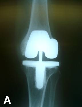
Figure 1A: These
are the anteroposterior radiographs of the left knee four years
after primary total knee arthroplasty. The
tibial component is in slight varus.
Figure 1B: These are the lateral radiographs of the
left knee four years after primary total knee arthroplasty.
An incomplete radiolucent line is noted under the anterior
flange of the femoral component.
The tibial component was noted to be in slight varus
alignment. A
three-phase technetium bone scan showed intense uptake around
the femoral component only. Blood indices and aspiration were within normal limits
with no evidence of infection. Revision knee surgery was undertaken, which revealed a
grossly loose femoral component and a well-fixed tibial
component. There were large contained bone defects involving
both femoral condyles. After
preparing the bone surfaces, a new constrained condylar femoral
component with distal and posterior augments was inserted along
with a diaphyseal press fit stem extension to engage the femoral
cortex. The core
implant was cemented distally, while the stem was press fit
proximally. The
patient had an uneventful recovery and is now without knee pain
at 18 months post revision.
Case Two
A
77 year-old female with a BMI of 30.9 presented three years following
right total knee replacement with worsening knee pain. She suffered from valgus osteoarthritis and had undergone
an Optetrak Non-Modular Constrained Condylar Knee replacement
without stem extensions (Exactech®,
Gainesville
,
FL
) because of inability to achieve coronal plane balance
following collateral ligament release. Initially, she had no instability, anatomic alignment of
5 degrees, and did well with no pain. She sustained a fall 6 months prior to presentation with
no significant injury but felt her pain had developed since
then. Physical
examination revealed a 5 degree flexion contracture and passive
flexion to 100 degrees. There
was a boggy effusion present and a suggestion of coronal plane
laxity. Tenderness
was elicited around the femoral component. Radiographs demonstrated a progressive radiolucency under
the anterior flange of the femoral component. Blood indices and aspiration were within normal limits
with no evidence of infection. A technetium bone scan demonstrated increased uptake
around the femoral component and a labeled white cell scan to
exclude infection was normal. Revision knee surgery was performed for persistent pain
and probable component loosening. The femoral component was found to be grossly loose. Significant contained femoral condylar defects from
toggling of the component were noted. Posterior and distal augments were used along with a
diaphyseal stem extension on a new constrained condylar femoral
component. Following
preparation of the bone the core implant was cemented distally
while the stem was press fit proximally. She made an uneventful post-operative recovery and is now
without knee pain 14 months years post revision surgery.
Case Three
A
72 year-old female with a BMI of 37.3 underwent a right Optetrak
Non-Modular Constrained Condylar Knee replacement without stem
extensions (Exactech®,
Gainesville
,
FL
) for valgus osteoarthritis. She had previously had a successful
Exactech® posterior stabilized left total knee replacement. The
patient did well initially with no instability. Two years after
her first index procedure, she reported that the right knee did
not feel like the left and had a tendency to give way. Radiolucency was noted behind the anterior femoral
flange. Blood
indices and aspiration were normal without any evidence of
infection. A three
phase technetium bone scan revealed increased uptake around the
femoral component, and an MRI suggested a large amount of
synovial debris and potential loosening of the patella without
loosening of femoral or tibial components. Because of persistence of symptoms consistent with
synovitis, an arthroscopic synovectomy was performed. The patients preoperative symptoms resolved but she
still reported achiness. Nine months later she had an episode of giving way and
fell. Radiographs
demonstrated translation and tilting of the femoral component. At the time of revision surgery, the femoral component
was grossly loose. Significant contained femoral condylar
defects from toggling of the component were present. All other
components were stable. Posterior and distal femoral augments
were used along with a diaphyseal stem extension on a new
constrained condylar femoral component. Following preparation of
the bone the core implant was cemented distally while the stem
was press fit proximally. She
made an uneventful post-operative recovery and the knee pain
resolved completely. She
is now 12 months post revision surgery.
Case Four
A
63 year-old male with a BMI of 23.7 presented with severe right
knee pain and a medial condyle fracture. Four years earlier he had undergone a bilateral knee
replacement with Optetrak Non-Modular Constrained Condylar Knee
replacement without stem extensions (Exactech®,
Gainesville
,
FL
) for varus osteoarthritis with lateral laxity. He did well initially with no instability, but three and
a half years post-operatively developed increasing pain and
difficulty walking. At
that time he was diagnosed with a soft tissue problem and
treated with anti-inflammatory medication. Physical examination revealed tenderness, crepitus with
range of motion, and a moderate effusion. Radiographs showed a fracture through the medial condyle
of the knee with displacement without any obvious trauma. Comparison to previous films indicated that the femoral
component had clearly shifted in position and loosened.
A CT
scan was also performed and demonstrated significant osteolysis
in the femur and some ostetolysis beneath the tibial base plate. Blood indicies and aspiration were normal with no signs
of infection. A
DePuy P.F.C. Sigma TC3 knee (DePuy
[Johnson and Johnson]. Warsaw, Indiana) with a rotating
platform base and a Synthes locking plate for the medial condyle
fracture (Synthes, West Chester, PA) were implanted with cement
because of the preference of this particular surgeon. The patient had an uneventful recovery and is currently
free of knee pain at 7 months post revision.
Discussion :
The
theoretical benefits of avoiding stem extensions with
constrained condylar knee replacements include prevention of fat
embolism from canal instrumentation, reduction in end of stem
pain, shorter operating times, less difficult surgery at
revision and reduced costs. Certain constrained condylar knees
have been developed that do not have a femoral stem extension so
as to minimize the violation of distal femoral metaphyseal bone. These relatively new implants are similar to a
posterior-stabilized femoral component except they have a wider
and deeper femoral box to accommodate the more conforming tibial
polyethylene spine. The
polyethylene in the Optetrak Non Modular Constrained implant is
machined from molded block material. The insert has 1.5° of medial-lateral motion and 2° internal-external rotation
before the femoral and tibial components begin to transfer and
share additional constraint. In all four of these patients Palacos cement (Zimmer®,
Warsaw
,
IN
) was used for fixation. These
three surgeons performed 125 primary total knee replacements
with this stemless constrained implant from
February 1, 2002
to
April 19, 2007
. These four
revisions are the only known cases of femoral component
loosening of implants inserted during this time period and
represent 3.2% of the total number of implants. Three of the
four patients described some traumatic event prior to loosening
and subsequent revision. This
compares to a revision rate of 6% for constrained condylar
implants using stem extensions (Genesis II, Smith and Nephew,
Memphis
,
TN
; Insall Burnstein CCK, Zimmer,
Warsaw
,
IN
; hinged implant, Biomet,
Warsaw
,
IN
) at our institution over a three year time period2. Specific data on revision rate for the Optetrak
Non-Modular Constrained Condylar Knee replacement with stem
extensions is unfortunately not currently available.
The theoretical risk of using constrained
condylar knee replacements without stem extensions is that the
interface stresses will be substantially higher and could lead
to early implant loosening. Both
Anderson
et al.1 and Nazarian et al.4 have reported
good intermediate term results when constrained devices were
used without stem extensions.
Anderson
et al.1 reported 49 patients (55 knees) who had
undergone primary total knee replacement with a constrained
condylar knee implant without stem extensions. At an average of 44.5 months follow-up, they found no
loosening, one dislocation, and one revision arthroscopy for
peripalettar fibrosis. Nazarian
et al.4 reported a rate of loosening of 10.1% for
revision total knee replacements using the stemless
Insall-Burstein constrained condylar knee implant at a mean
follow-up of 4.7 years. There
were 4 cases of tibial loosening and 2 cases of femoral
loosening in 55 knees and no significant differences in rate of
loosening between patients with implants with zero, one, or two
stems. A study by
Easley et al.3 found no loosening at 7.8 years when
stem extensions were used with these devices in spite of not
attempting to balance the ligaments. In a recent biomechanical study, Rawlinson et al.5
found that only specimens with reduced bone quality benefited
from the addition of a stem. Appropriate length and diameter were critical in
protecting the proximal tibia in these specimens. To our knowledge, the four patients included in this case
study are the only ones who underwent primary total knee
arthroplasty with this particular implant and were treated at
the same institution for loosening since February of 2002. This indicates a very low overall incidence of loosening.
These
cases also illustrate the fact that trauma, albeit minor,
following knee replacement with these types of implants should
be considered a causative risk factor for potential loosening.
It should be noted that nothing specific to this particular non
stemmed constrained condylar device was found to be contributory
to these clinical failures. While overt loosening may not be
noted clinically or even radiographically, further imaging
studies are warranted. In
each of the cases that we presented, the bone scan was useful to
demonstrate femoral component loosening. Patients that complain of persistent pain in the absence
of other definitive sources of pain should have a bone scan to
further evaluate the knee prosthesis for occult loosening. In addition, in every case presented the intraoperative
findings were much worse than anticipated, with severe
osteolysis, necessitating the use of stems and augments.
Until
more definitive evidence exists on the role of stems in
constrained condylar knees, we urge caution in the use of
non-modular or unstemmed constrained femoral components. With these stemless devices, the femoral component
appears more susceptible to early loosening than the tibial
component. In
addition, elderly patients with osteopenic bone may be more at
risk for such catastrophic early failure. Such patients in our practice now receive femoral and
tibial stem extensions to better distribute the stresses if a
constrained condylar implant is required.
Unfortunately,
the authors have noted a trend toward the more cavalier use of
stemless constrained femoral components and the lack of an
attempt to perform ligament balancing that may avoid these types
of prostheses. While
there may be a limited role for stemless constrained knee
prostheses in a select patient population (i.e. younger patients
with better bone quality), we recommend against the use of
unstemmed constrained devices and encourage surgeons to continue
to maintain the art of ligament balancing and insertion of a
traditional posterior stabilized devices if possible.
Reference :
-
Anderson
JA, Baldini A, MacDonald JH, Pellicci, PM, Sculco TP. Primary
Constrained Condylar Knee Arthroplasty without Stem Extension
for the Valgus Knee. Clin Orthop Relat Res 2006;442:199-203.
-
Boettner F, Laskin R, Windsor RE, Haas SB. Hybrid Component
Fixation in Revision Total Knee Arthroplasty. Clin Orthop Relat Res 2006;446:127-131.
-
Easley ME, Insall JN, Scuderi GR, Bullek DD. Primary Constrained
Condylar Knee Arthroplasty for the Arthritic Valgus Knee. Clin
Orthop Relat Res 2000;380:58-64.
-
Nazarian DG, Mehta S, Booth RE. A Comparison of Stemmed and
Unstemmed Components in Revision Knee Arthroplasty. Clin Orthop
Relat Res 2002;404:256-262. 2002
-
Rawlinson JJ, Peter LE,
Campbell
DA, Windsor R, Wright TM, Bartel DL. Cancellous Bone Strains
Indicate Efficacy of Stem Augmentation in Constrained Condylar
Knees. Clin Orthop Relat Res 2005;440:107-116.
|

