|
Abstract:
The true incidence of
extra articular synovial osteochondromatosis is unknown as a
proportion of patients will remain asymptomatic. It is thought
to be a rare entity with few previous case reports. We report on
a case in a 57 year old who despite florid clinical and
radiological findings had remained asymptomatic for many years.
J.Orthopaedics
2007;4(3)e20
Case
Report:
A 57 year old lumbarjack
was admitted to the orthopaedic ward after a heavy log had
fallen onto the lateral aspect of his left leg. He presented
with pain and swelling over the proximal aspect of the left
tibia which was associated with ankle weakness. There were no
other further injuries.
His past history
included small vessel vasculitis. His current medication
included omeprazole 10mg od, prednisalone 5mg od and
azathioprine 50mg od. He also reported a non-progressive
swelling located over the lateral aspect of the left knee. This
swelling had been present for many years although had not caused
him significant symptoms.
On examination he was
found to have no other injuries. Examination of the left leg
revealed bruising and tenderness over the proximal fibula. This
was association with the long-standing firm smooth nodular
swelling over the lateral aspect of the left knee. The swelling
was non-tender and measured 7 centimetres by 7 centimetres
(Figure 1). The overlying skin was freely mobile with no
tethering. He had normal knee range of motion with no effusion
and the knee was stable. Neurological assessment revealed grade
1 MRC power in EHL and ankle dorsiflexors. Sensation was reduced
in the distribution of the common peroneal nerve.
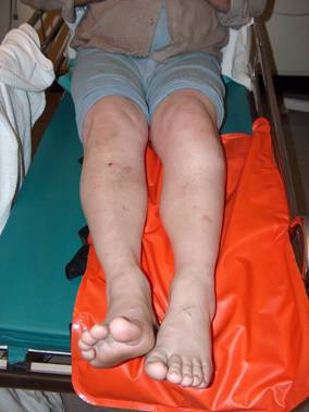
Figure 1: Clinical
photograph showing mass proximal left tibia and foot drop
Plain radiographs showed
no fracture but an incidental finding of multiple calcific
nodules which appeared to represent extra-articular synovial
chondromatosis (Figures 2 and 3).
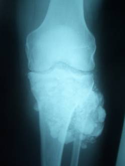
Figure
2: Plain antero-posterior radiograph
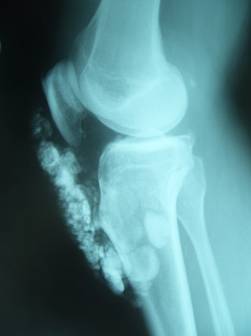
Figure
3: Plain lateral radiograph
He was managed with
physiotherapy and a foot drop splint. At two week follow up his
symptoms had almost completely resolved and he was found to have
normal neurological function.
Discussion:
Synovial
osteochondromatosis (also called synovial chondromatosis,
synovial chondrometaplasia, synovial chondrosis and synovial
chondromata) is a benign synovial neoplasm and most commonly is
monoarticular. This condition was originally described by Freund
1 in 1937 and is believed to be the result of
synovial metaplasia. Further studies suggest that the synovial
connective tissue undergoes cartilaginous metaplasia 2 .
These cartilage nodules may then be converted to bone
through the process of encondral ossification.
This is a rare condition
which may occur intararticularly or extraarticularly. It can
occur idiopathically (primary) or as a result of localised joint
soft tissue irritation (secondary). It is most often seen in its
intra-articular form in the adult population but has also been
reported in children 3. Within joints these nodules
are thought to form from articular synovial membrane while
extraarticular nodules originate from bursal or tendon sheath
synovium.
Osteochondromatosis is
thought to be a benign condition but is often frequently
recurrent and usually gives symptoms in the form of joint pain,
swelling and osteoarthritic changes related to a mass effect 3.
Malignant transformation of intraarticular synovial
osteochondromatosis can occur but is rarely reported 4,5.
Furthermore, to our knowledge, there has been only one report of
possible malignant transformation with the extra-articular form 6.
If the osteochondromatosis lesion is adjacent to extraarticular
bone it is important to rule out other conditions such as
synovial sarcoma or periosteal chondroma7.
The true incidence of
this condition is unknown; most of the literature relates to the
more common form of intraarticular osteochondromatosis. A
thorough medical history and clinical examination should be
obtained, followed by plain radiographs and MRI scanning. In
this case the patient had been asymptomatic for many years and
the radiological findings were incidental. He did not wish
further investigation or treatment and had returned to work with
normal foot function within ten days of his injury.
Reference :
- Freund
E. Chondrosarcomas of the joints. Arch Surg. 1937;34:670
- Jaffe
HL. Synovial chondromatosis and other articular tumours. In:
Tumors and tumorous Conditions of the Bones and Joints.
Philadelphia
: Lea and Febiger, 1958:566-567
- Ko
E, Mortimer E, Fraire A. Extraarticular synovial
chondromatosis: review of epidemiology, imaging studies,
microscopy and pathogenesis, with a report of an additional
case in a child. Int J Surg Pathol 12(3):273-280, 2004
- Taconis
WK, van der Heul RO, Taminiau AM. Synovial chondrosarcoma:
Report of a case and review of the literature. Skeletal
Radiol 26:682-685, 1997
-
Blokx
WA
, Rasing LA, Veth RP, Pruszczynski M.
Late malignant transformation of biopsy proven benign
synovial chondromatosis: An unexpected pitfall.
Histopathology 36:564-566, 2000
- Kaiser
TE, Ivins JC, Unni KK. Malignant transformation of
extra-articular synovial chondromatosis: Report of a case.
Skeletal Radiol 5:223-226, 1980
- Sim
FH,
Dahlin
DC
, Ivins JC. Extra-articular synovial chondromatosis. J Bone
Joint Surg Am 59:492-495, 1977
|

