|
Abstract
We describe a case of Ulnar Dimelia commonly called Mirror
hand. This is an exceedingly rare congenital anomaly of the upper
limb. In this case we found a complete dislocation of the
shoulder with a limited range of movement. No other facial or
internal organ malformation, the patient has complete absence of
the Radius including the capitalum.
J.Orthopaedics 2006;3(4)e15
Case Report
A two years old female patient presented
with left upper limb deformity since birth involving hand,
wrist, elbow and shoulder impeding the normal function of the
limb. She is the second child of the family. There was no
similar condition among the family or relatives.
On examination, the patient was well built, with good
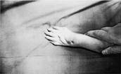
mentality. Localy, the left hand has seven fingers, six of them
are well formed but the seventh is small and rudimentary. All
fingers lie in the same plane with slight apposition between the
two halves. There is no thumb. The fingers are somewhat flexed,
and the hand as a whole is usually radialy deviated at the wrist
and dropped and cannot be extended
The elbow is stiff in extension and the
arm is short. Pronation and supination were limited. As to the
shoulder, there is loss of the normal contour, with prominence
of the tip of the acromian, all shoulder movements were grossly
restricted especially abduction , with the humeral head can be
felt in the axilla lying against the ribs.
A picture showing the absence of the
thumb. The fingers are all in one line
X- Ray showed The ulna
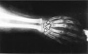 and the ulnar carpal bones are completely
duplicated, the scaphoid and trapezium are replaced, and distal
ulnar epiphysis is broadened. and the ulnar carpal bones are completely
duplicated, the scaphoid and trapezium are replaced, and distal
ulnar epiphysis is broadened.
At the elbow, each of the duplicated ulnae articulates with the
distal humerus separately and they tend to face each other
.There is no capitalum on the distal humerus. At the shoulder,
it is dislocated with the glenoid.hypoplasia of the Scapula.
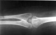
(Fig.3) an Ap view of the forearm
Discussion :
Ulnar Dimelia occurs as radial and ulnar clusters of fingers in
the same hand that are near images of each other. It is
considered aduplication phenomenon of substitution of the
radial components as well,
The understanding of the embryology of mirror image deformities
has increased dramatically since the early work of Saunders and
Gasseling, who first produced mirror image digit duplications in
chicks by grafting a small piece of posterior border mesoderm
into an anterior position(9).
It is now understood that this zone of polarizing activity
expresses the gene sonic hedgehog, which regulates limb
patterning on an anteroposterior axis(5,6). Ectopic expression
of the zone of polarizing activity cells or the sonic hedgehog
gene can therefore produce mirror image deformities(1).
More recently, secondary signaling molecules such as bone
morphogenic proteins and certain Hox genes have been implicated
in the embryogenesis of mirror image deformities(6). Hox genes
encode positional information during embryogenesis. Hox b-8 is
thought to be important in the specification of the zone of
polarizing activity cell positioning, ectopic expression of
which has experimentally resulted in mirror image
duplication(3,6) Retinoids seem to regulate the expression of
Hox genes(3).
Others may argue that Ulnar Dimelia is not as easily classified
as pure duplication (11, 8). This indicates that the Radius was
absent and the ulna noticed in place of the radius. Her the
archipterygeal theory could be taken into consideration. It is
stated that due to reduplication the main stem resulted in the
development of the double ulna(13).
Approximately 70cases have been reported, 4 of which with
shoulder dislocation including our case(7) . The largest
reported series is that of Harrison, Pearson and Roaf in which
they described the deformity in three patients (10), its
occurrence is usually sporadic (10).
Ulnar dimelia is usually associated with some degree of
hypoplasia of the arm and scapula which is present in this case,
and sometimes with fibular dimelia and absent tibia which is not
in our case. However: a multimodality imaging approach,
exploring the various aspects of the malformation is mandatory
to help the surgeon in order to obtain a functional and
aesthetic upper limb after complex surgical procedures taking
into account the various aspects of the malformation(7).The
reconstructive procedures are described toward restoring elbow
flexion, forearm supination (by excision of the proximal part of
one of the Ulnae ), wrist extension and policization to allow
apposition (4).
Reference :
-
Viljoen, D. L., and Kidson, S. H. Mirror polydactyly:
Pathogenesis based on a morphogen gradient theory. Am. J. Med.
Genet. 35:229, 1990.
-
Sinha, D.N. Possibility of Cell Death Induced Skeletal
Malformations Of The Upper Limb J Anat. Soc. India 49(2) 158-160
(2000) Vol. 49, No. 2, December, 2000.
-
Hatchwell, E., and Dennis, N. Mirror hands and feet: A further
case of Laurin-Sandrow syndrome. J. Med. Genet. 33:426, 1996.
-
Lister G: Wrist and Hand : Congenital Anomalies And Pediatric
Reconstruction .In: Orthopedic Knowledge Update 4, Upper
Extremity ,Chapter 30. p 178- 180.
-
Hersh, J. H., Dela Cruz, T. V., Pietrantoni, M., et al. Mirror
image duplication of the hands and feet: Report of a sporadic
case with multiple congenital anomalies. Am. J. Med. Genet.
59:341, 1995
-
Al-Qattan, M. M., Al-Thunayan, A., De Cordier, M., Nandagopal,
N., and Pitkanen, J. Classification of the mirror hand: Multiple
hand spectrum. J. Hand Surg. (Br.) 23:534, 1998.
-
Radiol. J Ulnar dimelia:
imaging modalities and surgical implications 2000
Mar.;81(3):219-22.
-
Chinegwundoh JO,
Gupta M,
Scott WA Ulnar dimelia. Is it a
true duplication of the ulna?
J Hand Surg. [Br]. 1997
Feb;22(1):77
-
Saunders, J. W., and Gasseling, M. T. EctodermalMesenchymal
Interactions in the Origin of Limb Symmetry. In R. Fleishnajer
and R. E. Billingham (Eds.), EpithelialMesenchymal
Interactions. Baltimore: Williams & Wilkins, 1968. Pp. 7897.
-
Harrison RG,Pearson MA, Roaf 1960 J.B.J.s , 42-B: 549 .
-
Mark T. Jobe and Phillip E. Wright II . Congenital Anomalies of
Hand. In: S .
-
Terry Canale, editor. Campells Operative Orthopaedics,9th.
Edition, ,1998. Vol.4 p.3792.
|

