|
Abstract:
Background: Biodegradable implants after their successful
worldwide usage have recently entered Indian orthopaedics. Since
then there are only handful comments on their utilisation for
fracture fixation in this developing country. This study has
evaluated results of biodegradable implant fixation in commonly
occuring intraarticular fractures around elbow and ankle joint.
Materials and Methods: A prospective study was undertaken to
evaluate functional outcomes and complications of fractures
stabilized with biodegradable (PLLA)
implants in Indian set up. Thirty two patients including
twenty childrens (< 14 yrs) and twelve adults of closed
intraarticular fractures around elbow and ankle undergone
fixation with biodegradable implants. Results were evaluated
after a minimum follow up period of six months. Mayos scoring
system was used for elbow cases and Olerude-Molander scoring was
used for ankle cases. Functional outcome and complications were
noted.
Results: Fracture union and
functional outcome at six months was satisfactory among all
cases. Two elbow cases showed inflammatory swelling at
operative site at fourteenth week followup. Two showed
extraosseous calcification and one had malunion. Five patients
suffered stiffness of elbow joint for some time during their
followup. One patient of ankle fracture had displacement and
infection. Other four cases had stiffness. No any child among
cases followed more than thirtytwo weeks showed growth
disturbance or abnormal carrying angle. No patient of either
elbow or ankle suffered sterile abscess or sinus.
Conclusion: Biodegradable implants could be advantageous in
properly selected intraarticular fractures around elbow joint,
especialy in children. They are no better than metal implants
for fractures around ankle joint because of delayed mobilization
and associated stiffness.
J.Orthopaedics 2010;7(4)e7
Keywords:
Biodegradable Implant; Intraaritcular Fixation; PLLA.
Introduction:
The
internal fixation devices used in fracture repair serve to hold
the fractures in proper alignment and apposition till the
fracture unites, after which they do not serve any function.
Although protocol for removal of
these
hardware varies greatly among different institutes, from regular
removal to only from those with symptoms and complications.
These retained metallic devices proved to have some adverse
effects, the most important of which are osteopenia of cortical
bone induced by stress protection1, others include
corrosion and sensitisation reactions with some alloys2,3.
These seem to be of minor importance
when fractures are confined to cancellous bone and modern
biologically inert implants (Titanium) are used. However, there
are always patients who request removal of implant because of
chronic irritation of the surrounding soft tissues by prominent
hardware4. This removal surgery besides being an
economic burden, pushes the patient through same physical and
psychological stresses of that of initial surgery. In this
scenario bioabsorbable (PLLA)
implant is a logical way to proceed for fracture fixation.
Displaced malleolar fractures are common injuries, and the
functional results are strongly correlated with anatomical
reduction and internal fixation24.
Fractures of the lateral condyle of the humerus are common in
childrens and usually treated by open reduction and internal
fixation to ensure proper union and normal growth23.
These were the reasons to include these fractures in this study.
Many
studies from developed world in previous four decades proved the efficacy of these
devices
as similar as that of metal
implants4,5,6,9,12,14,19,21,22. Varying opinions had
come up among their users in trauma surgery in relevance to
advantages over conventional metal implants4, 5,6,8,9.
Now these implants have been
introduced in the Indian orthopaedics18.
Little
has been published about this topic in orthopaedic literature in
India 7,18. Very few Indian studies till date, as
per our knowledge has evaluated these implants for
intraarticular fracture fixation. This study reports thirtytwo
patients of commonly occurring intraarticular fractures around
ankle and elbow, fixed with biodegradable implants in Indian
setup is unique in its kind.
Materials
and Methods:
Materials & Methods
Study Design
A prospective trial was carried out for Biodegradable i.e.
Poly-L-Lactic Acid (PLLA) implant [INION, Finland]
in the internal fixation of intra articular fractures
around elbow and ankle joints after approval by Hospital ethics
committee. During the study period (July 2006 to November 2009)
thirty eight patients with intraarticular fractures of elbow and
ankle were enrolled in this trial. The patients were explained
about the advantages, known risks and cost factors associated
with biodegradable implants. Four patients could not afford for
this implant and two patients didnt returned for first
followup also, so excluded from final evaluation. This left with
thirty two patients including twenty childrens (<14 years of
age) and twelve adults.
Table. 1 : Patient Particulars
|
|
|
|
Number of cases |
Lateral condyle humerus |
18 |
|
Capitulum |
6 |
|
Medial malleolus |
4 |
|
Bimalleolar |
2 |
|
Trimalleolar |
2 |
|
Sex (Male : Female) |
13:3 |
|
Mean age in years (range) |
20.68 (3 to 58) |
|
Average period of follow-up (Months) |
27.1 |
|
Average days of hospital stay |
5.2 |
Informed consent was given by the parents in case of minor
patients and by patients in case of adults to be included in
this study. Mandatory minimum follow up period was 6 months.
Patients having closed
intraaritcular fractures around elbow or ankle joints with
displacement of one millimeter or more needing open reduction
and internal fixation were included in this study. Medically
unfit, chronic alcoholics, psychiatric and uncooperative
patients were excluded.
Table. 2: Fracture Patterns
|
Fracture |
|
|
|
Male |
Female |
|
|
Lateral condyle Humerus |
Type 1 |
|
|
|
Type 2 |
16 |
2 |
|
Capitulum |
Type 1 |
4 |
2 |
|
Type 2 |
|
|
|
Medial Malleolus |
Oblicque |
4 |
|
|
Trasverse |
|
|
|
Bimalleolar |
2 |
|
|
|
Trimalleolar (Cottons) |
|
2 |
|
Operative Technique
Under general or regional anesthesia, fractures were exposed
using standard approaches. Procedures were performed in
bloodless field under control of tourniquet. Procedure for
placement of biodegradable screw is essentially same as that of
its metal counterpart (i.e. making drill hole, tapping and
placing the screw) except that the screw driver used for PLLA
screws are star tipped and torque-limiting, to avoid damage to
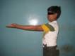
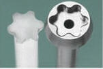
screw head. (Fig.1)
Fig.1:
Star tipped Torque Limiting screw driver used for Biodegrdable
Screw
For lateral condyle of
humerus, Kochers lateral approach was used, each fracture was
reduced and held with bone clamps. Fine Kirschner wires were
also used whenever necessary to provide temporary fixation. In
initial two cases of lateral condyle fracture of humerus, PLLA
pins were used but as the stability was not adequate, additional
metallic K-wire in one case and PLLA screw in another case was
used to supplement the fixation. In remaining all lateral
condyle and capitulum fracture cases PLLA screw was used.
In ankle fractures, medial malleolus was fixed with a 4.5mm
biodegradable (PLLA)
screw and fibula was fixed with a biodegradable plate.
Technique of application of biodegradable malleolar screw and
fibular plate was same as that of routinely used metal implants,
except that PLLA fibular plate is malleable at specific
temparature and can be contoured by molding it after heating in
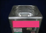
thermo water bath at 75oC (Fig:2).
Figure.2: Thermo-water bath Used to heat fibular plate to make
it malleable
Patients were followed post operatively at 2 wks, 6 wks, 3
months and 6 months. Patients were given appropriate supportive
slabs until suture removal after 2wks of surgery. Assisted
active mobilization of elbow
cases was allowed after suture removal. Ankle cases given
further four weeks of splintage. Partial weight bearing was
allowed after six weeks in
ankle cases. Full weight bearing was allowed after three
months. During this period patients were evaluated clinically,
subjectively and radiologicaly for functional outcome. Standard
scoring systems i.e. Mayos scoring system for elbow joint and
Olerud Molanders scoring for ankle joint were used. Any
complications that were encountered were noted.
Results :
In the elbow fractures, the strength of fixation achieved at
surgery was satisfactory. The wounds healed normally in all
patients. Twelve patients (50%) of twenty four elbow cases
showed excellent functional score (Fig.3), eight patients
(33.3%) had good functional score, three patients (12.5%) had
fair score and one patient
showed poor result. Mayos elbow score was used to evaluate
functional outcome.
Fig.3 : A Case of fracture lateral condyle humerus with
dislocation elbow
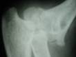 
 
a)PreOp X ray b) At 18 months Follw-up
Discussion :
There was no evidence of
infection or loss of fracture fixation in any of the children
but five patients suffered stiff elbow for some time during
their follow up period. A case of fracture lateral condyle
humerus had malunion (Fig.5b). review showed that all patients had full elbow
movement and normal function. Out of twenty childrens the
thirteen casese were followed for more than 32 months.
None of these cases showed any evidence of abnormality in
physeal growth or carrying angle.
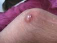
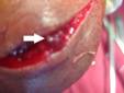
Fig.5 : Complications Extraosseous Calcification & Malunion
   
a.Two different patients showing Extraosseous calcification(
Arrow)
 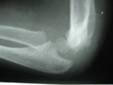
b. A case of Malunion Lateral condyle Humerus
Clinical results among
ankle cases operated with these implants were not as encouraging
as that of elbow. Two out of eight ankle cases showed excellent
result, one case showed good result, four showed
fair results and one showed
poor result. Four out of eight ankle patients had stiffnes. A
patient of trimalleolar fracture operated initially in this
series got displacement of fragments, may be due to over
reliance on stability of fixation with these implants and
partial weight bearing at six weeks. Same patient suffered
infection also which further deferred mobilisation. So in later
ankle cases we have extended splintage up to six weeks.
No patient in this
series suffered frank sterile abscess or sinus, which was common
occurrence with first generation of biodegradable implants.
There was no periprosthetic osteolysis and deep infection
observed. Although occurrence of inflammatory swelling at
operative site in two lateral condyle humerus patients needs
further investigation. This complication is not common with
Poly - L Lactic acid implants.
Discussion
Research for
use of bioabsorbable implants in the repair of bone tissue began
in the late 1970s; the initial studies were performed in the
field of maxillofacial and mandibular surgery 28,29,30.
Initial implant material was poor in strength which was not
sufficient for orthopaedic application. After introduction of
self reinforcing technique in manufacturing of theses materials,
their use expanded to be included in orthopaedics 4 .
Since then, they
have been reported to be used in internal fixation of fractures
of the ankle (Bostmann et al l989, 1990)8,10, elbow (Hirvensalo
et al 1988b)26 and distal radius (Hoffmann et al
1989)27, and to secure osteotomies of metatarsals (Hirvensalo
et al l988a)25 . In these strengthened first
generation implants, PGA
(Polyglycolic acid) was the main constitute. Later this material
found to have rapid degradation rate leading to premature
decrease in fracture stability and formation of typical sterile
abscess and sinus.
Presently in use second
generation biodegradable implants are copolymers made up of
PLA(Poly Lactic Acid) as their main core element, with
initial molecular weight of 1,80,000
to 5,30,000 and melting point of 174oC . Particularly
its levo isomer (PLLA) has long degradation time i.e. two
to five years, providing enough strength and stability for
enough period 7.
P. G. Hope et al6
in their series of twenty-four patients compared
biodegradable implants with metallic Kirschner wires in
displaced fractures about elbow. Fracture union with full
function occurred in all cases within six months. Kirschner
wires caused problems including infection, soft-tissue
ossification and required removal under general anaesthesia.
Results of present study are also not different from this study
except that biodegradable screws were used rather than pins. In
initial two cases stability provided by pins was not adequate
for complete reliance and required supplementation with metal
implants. Advantage of avoidance of second surgery for removal
proved definitely a major factor for their use in children with
fracture lateral condyle humerus in whom general anaesthesia is
necessary.
In addition, lesser total days of
hospital stay (including hospitalisation during implant removal
surgery) should also be taken as a positive factor in favor of
biodegradable implants.
O Bostman et al
in his series of fifty six patients of displaced malleolar
fractures 8 and in their series of 102 patients of
unimalleolar or bimalleolar fracture11, found no
change in the ability to participate in sports and other
physical activities at one year follow-up.
Ankle cases however in
present study did not perform according to their elbow
counterparts. This comparison although is irrational keeping in
mind different anatomical and biomechanical properties of bones
at these two different sites. Moreover difference in age group
of occurrence of fractures, (i.e. lateral condyle humerus
fracture mainly occcurs in children and ankle fracture is the
fracture of adults) is another major factor for varied results
among performance of this implant at these two different sites.
The results showed ankle patients needed prolonged
immobilization and developed subsequent stiffness. The worst
part of present series was a case of Cottons fracture treated
with biodegradable implant, which underwent implant displacement
and infection after six weeks. Although the number of ankle
cases was limited to draw any firm conclusion, but facts were
not in favor of use of biodegradable implants in this weight
bearing joint.
Regular use of
biodegradable implant in developing countries has its own
limitations. First and foremost is the cost of implant, which is
at eight times more than that of Indian made stainless steel
implants. Next is the availability of implants. Only a few of
big cities in India have access to these devices. Familiarity
and confidence of surgeons with these devices is also an issue
yet to be solved. As biomechanical properties of these implants
differ with that of metal, and a different instrumentation kit
is provided with the set of implants, operating with these
devices has a learning curve. All of four senior surgeons of
our institute having experience of operating with these implants
didnt find themselves comfortable with biodegrdable pins in
regard to purchase of bone and strength of stability provided by
these devices. So to take advantage of properties of these
devices in, they should be used for proper indications and in
hands of experienced person who are well versed with their use.
Despite these limitations, , patients are turning up to
opt for these implants, may be due to psychological impact of
the fact that, these implants dissolves in body and doesnt need
another surgery merely to remove it. But their cost
effectiveness over metal implants in established indications is
a matter of research. To study their efficacy and feasibility in
India, further long-term follow up and a larger study design is
required.
Conclusion:
This study has shown
that biodegradable implants are a promising modality of
treatment for intraarticular fractures around elbow joint,
especially in children in whom general anaesthesia is often
required for removal of metal implants. Their use in
intraarticular ankle fractures is not as encouraging and
advantageous as conventionally used metal implants. So this
study doesnt recommend their use in this weight bearing joint.
Cost, availability and familiarity with these implants are other
issues associated with these devices that need further
evaluation.
Reference:
-
Laftman P, Nilsson OS (1989): Stress shielding by rigid
fixation studied in osteotomized rabbit tibiae. Acta Orthop
Scand 60: 718-722.
-
Benson MKD, Goodwin PG, Brostoff J (1975) : Metal sensitivity
in patients with joint replacement arthroplasties. Br Med J
4:374-375
-
Barranco VP, Solomon H (1972): Eczematous dermatitis from
Nickel. JAMA 220:1244
-
O.M. Bostman, M.D., Current Concepts Review Absorbable
implants for the fixation of fractures.
J Bone Joint Surg (Am), Vol. 73-A, 148-153, January
1991.
-
Bostman O.; Vainionpaa, S.; Hirvensalo, E.; Makela. A.;
Vihtonen, K.; Tormala. P.; and Rokkanen, P.: Biodegradable
Internal Fixation for Malleolar Fractures. A Prospective
Randomised Trial. J. Bone and Joint Surg., 69-B(4): 615-619,
1987.
-
P. G. Hope, D. M. Williamson, C. J. Coates, W. G. Cole
Biodegradable pin fixation of elbow
fractures in children A Randomised Trial:Jbjs(Br),73-B,965-8,1991
-
MS Dhillon,Lokesh AV : Current concept review
Bioabsorbable implants in orthopaedics. Indian Journal of
Orthopaedics Oct 2006; vol 40 :p. 205-209
-
O.Bostman, M.D., E. Hirvensalom,. D., S. Vainionpaa, M.D.,
Makela,M.D.,K. Vihtonenm,.D., P. Tormala, Ph.D., and P.
Rokkanenm,.D Ankle Fractures Treated Using Biodegradable
Internal Fixation: J. Clinical Orthop & Related Research, No
238, Jan 1989.
-
Bostman, E. A. Makela, P. Tormala,P. Rokkanen
Transphyseal Fracture Fixation using Biodegradable
Pins:Jbjs(Br), 71-B,706-7,1989.
-
O. Bostman, E. Hirvensalo, J. Makinen,P. Rokkanen Foreign-body
reactions to Fractture Fixation
Implants of Biodegradable Synthetic Polymers:Jbjs(Br),72-B,592-6,1990
-
Seppo Santavirta, Yrjo T. Konttinen, Tomoyuki Saito, Mats
Gronblad, Esa Partio, Pertti Kemppinen,
Pentti Rokkanen: Immune response to Polyglycolic Acid
Implants:Jbjs(Br),72-B,597-600,1990
-
P. P. Casteleyn, F. Handelberg, P. Haentjens, Biodegradable
rods versus kirschner wire
fixation of wrist fractures a randomised Trial: J Bone
Joint Surg [Br] 74-B :858-61,1992.
-
H. Pihlajamaki, 0. Bostman, E. Hirvensalo, P. Tormala,P.
Rokkanen: Absorbable pins of self-reinforced
poly-l-lactic acid for fixation of Fractures and Osteotomies:
J Bone Joint Surg [Br] 74-B : 853-7.199
-
R. K. Fraser, W. G. Cole Osteolysis After Biodegradable Pin
Fixation Of Fractures In Children: J Bone
Joint Surg [Br] 74-B : 929-30, 1992
-
Bostman. M.D. U. Paivarinta, M.D., E. Partio. M.D., J.
Vasenius. M.D. M. Manninen.
M.D. And P.
Rokkanen. M.D.: Degradation And Tissue Replacement Of An
Absorbable Polyglycolide Screw In The Fixation Of Rabbit
Femoral Osteotomies. Jbjs (Br)Vol
74-A,No.7,1021-1031,Aug 1992
-
G. O. Hofman: Biodegradable implants in traumatology: a review
on the state-of-art; Arch Orthop Trauma Surg114:123-132, 1995
-
M. Fuchs · G. Köster · T. Krause · H.-A. Merten
·
A. Schmid :Degradation
of and intraosseous reactions
to biodegradable poly-L-lactide screws, a study in minipigs:
Arch Orthop Trauma Surg, 118 :140144, 1998
-
M.S. Dhillon, S.Prabhakar, C.Prasanna:Preliminary experience
with biodegradable implants for fracture fixation: Indian
Journal Orthopaedics,Vol-42, Issue 3, 319-22, Jul- Sept 2008
-
T. Juutilainen, H. Patiala, P.Rokkanen, P.
Tormala:Biodegradable wire fixation in olecranon and patella
fractures combined with biodegradable screws or plugs and
compared with metallic fixation, Arch Orthop Trauma Surg,114;
319-323, 1995
-
Evers, B; Becker, H-P; Gerngross,H: Long- term results After
Use of Biodegradable Implants (Biofix(r)) in Orthopaedic
Surgery; JBJS Br Pg 237,Vol 79-B (2S), Suppl Jul 1997
-
Claes, L.; Rehm, K.; Helling, H. J.; Hutmacher, D.: The
fixation of small bony fragments with a new biodegradable
implant: Orthopaedic Proceedings: European Orthopaedic
Research Society: Munich, Germany - July 2-3, 1995
-
Ranko Bilic´, Robert Kolundzˇic´, and Darko Anticˇevic´:
Absorbable Implants
in Surgical Correction of a Capitellar Malunion in an
11-year-old A Case Report,
J Orthop Trauma 20:6669;2006.
-
Rang M.
Childrens fractures.
2nd ed. Philadelphia etc: JB Lippincott Company, 1983.
-
Tunturi T, Kemppalnen K, Patialal H, Suokas M, Tamminen 0,
Rokkanen P. :Importance of anatomical reduction for subjective
recovery after ankle fracture. Acta Orthop Scam: l983;54:64.
-
Hirvensalo E, Bostman 0, Vainionpaa S, Tormala, Rokkanen P.
Biodegradable fixation in chevron osteotomy for hallux valgus.
Acta Orthop Scand
l988a; Suppl 227 :71.
-
Hirvensalo E, Boatman 0, Vainionpaa, Tormala, Rokkanen P.
Biodegradable fixation in intraarticular fractures of the
elbow joint. Acta Orthop Scand 1988b; Suppl 227 :78-9.
-
Hoffmann R, Krettek C, Haas N, Tscheme H. The distal radius
fracture: operative stabilization with biodegradable fracture
of the pins (Biofix). Unfa//chirurg 1989; 92:430-4.
-
Cutright, D.E., Hunsunk E. E. The Repair of Fractures Of
Orbital Floor Using Biodegradable Polylactic Acid.
Oral Surg. 33; 28-32, 1972
-
Getter Lee, Cutright, D.E.,Bhaskar S.N., AugsburgJ.K.: A
Biodegradable Intraosseous Appliance In The Treatment of
Mandibular Fractures.
J Oral Surg 30: 344-348,1972
-
Miller,
R. A. Brady, J. M.; and Cutright, D. E. : Degradation Rates of
Oral Resorbable Implants (Polylactates and Polyglycolates):
Rate
Modification with Changes in PLA/PGA
Copolymer Ratios. J. Biomed. Mater. Res. 11: 71 1-719,
1977.
|