|
Abstract:
Aseptic necrosis of the lunate bone in its advanced stage
presents a large joint destruction. His treatment consists in
total wrist fusion. We report a patient with Kienböck, grade IV
of Litchmanns classification, who underwent proximal row
carpectomy and placement of a Resurfacing Capitate Pyrocarbon
Implant (RCPI).
J.Orthopaedics 2010;7(3)e10
Keywords:
Kienböck; asseptic necrosis of the lunate proximal carpectomy;
RCPI; wrist arthroplasty.
Introduction:
Although it was described more than 150 years ago, the etiology
of the Kienböck disease is unknown.
Aseptic
necrosis of the lunate is studied at the Federal University of
São Paulo for over 20 years. The
relationship between the incidence and risk factors for the
Kienböck disease are difficult to establish, because it is a
rare condition. It usually affects adults, being more common
among men (1,2).
The optimal treatment has not been established yet and, varies
according to the authors. The advanced stages IIIB and IV are
characterized by progressive carpal collapse, change of carpal
kinematics, fragmentation of the lunate and secondary
osteoarthritis. The proposed treatments are controversial and
remain a therapeutic challenge. At this stage, the lunate is not
amenable to surgery reconstitution; it is only possible salvage
surgery. Several surgical techniques have been proposed, like
denervation of the wrist, lunate resection and tendon
interposition, revascularization of the lunate, shortening
osteotomy of the radius, osteotomy for lengthening of the ulna,
lowering of the capitate osteotomy, parcial or total wrist
fusion, total wrist arthroplasty, carpectomia proximal and,
lunate replacement by silicone prosthesis. There is no strong
evidence of the superiority of one procedure over another
(3,4,5).
The goal of our work was to report a patient with grade IV
Kienböck that in lieu of the total wrist arthrodesis, underwent
surgery for placement of a capitate head prosthesis made of
pyrocarbon (RCPI).
Case Description
JMA, male, 38 years, wall painter, born and raised in São Paulo,
Brazil. Patient reported complaint of pain in the left fist,
which begun insidiously six months ago. The pain was accompanied
by progressive loss of range of active and passive movements (20
° - 20 ° of flexion-extension).
Examinations of X-rays and MRI have confirmed the necrosis of
the lunate (Kienböck) grade IV of Litchmanns classification
(Figure 1).
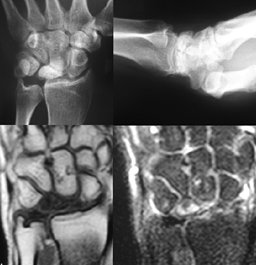
Figure 1:
Image of X-rays and MRI showed the involvement of the
radiocarpal and medio-carpal joint.
Surgical Technique
The patient in the supine position was subjected to anesthesia
type brachial plexus block.
A dorsal and radial incision in the left wrist was performed,
opening between the second and third tunnel extensor.the capsule
was opened by the technique in ">" to preserve the ligaments.
During resection of the first row of the carpus and
radial styloid osteotomy, was
possible to observe the articular cartilage lesions of the head
of the capitate and lunate fossa on the radio. Then carefully,
all articular cartilage of the head of the capitate bone was
removed with minimal resection of the proximal part (convex
shape) in order to expose the cancellous bone. If possible, the
resection should be parallel to the radius distal side. A sharp
awl is used to prepare the hole and locate the central aspect of
the medullary canal of the capitate. Preparation of the capitate
was made, using the broaches (and eventually the impactor/
extractor screwed on their extremity), starting with the
smallest size. A guide mark shows the dorsal side on the broach
to prepare the capitate with the correct orientation (Figure 2).
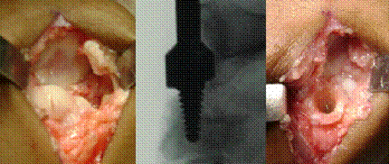
Figure 2:
Injury in the cartilage surface of the radio. Radioscopy showing
the position of the cutter within the capitate. Canal formed in
the capitate.
Next we tested the evidence of the prosthesis to check what size
was more appropriate. The adequate size and position were
confirmed by fluoroscopy. Then, we did the replacement of the
trial implant by the corresponding pyrocarbon implant with a
plastic tong to prevent any injury to the prosthesis. The
procedure of closure was normal and no additional stabilization
technique was required. The wrist was splinted for 3 weeks and
unrestricted active range of motion was allowed after eight
weeks with strengthening started after 12 weeks.
In evaluating the patient in the first year after surgery, he
found himself satisfied with the surgery and had returned to his
job. He complained of pain in small intensity (intensity in an
analog scale of pain) to extreme efforts. Inspection showed a
hypertrophic scar on the dorsum of the wrist and the presence of
palmar callosities. The presence of callosities demonstrated
that the patient had a heavy manual work activity (Figure 3).
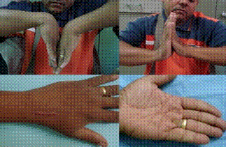
Figure
3: Clinical appearance at 1 year of follow-up. Range of
active flexion and extension movements of the wrist.
Hypertrophic scar. Palmar callosities.
The active and passive joint range of flexion increased from 20
° in pre to 60° postoperative and remained in the extension 20°.
In the X-ray image, the prosthesis was observed to be in
position equal to the intraoperative period. A comparison of the
postoperative first week and one year examination, show the
latter a halo of bone reabsorption between implant and bone
(Figure 4).
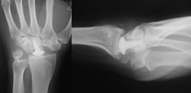
Figure 4:
X-Ray showing position of the prosthesis with 1 year after the
surgery.
Discussion :
In a recent
publication, Lumsden et al (2008) showed good results after 15
years following proximal row carpectomy in stages III. Also in
2008, Stern and Croog (2008) showed that the three patients who
underwent proximal row carpectomy, required reintervention for
total wrist fusion, two patients were grade IV. We believe that
to Grade IV would be the best indication to surgical total wrist
arthrodesis, but this is not a surgery without complications and
has the disadvantage of taking the whole movement of the wrist.
The total wrist prosthesis has a very high cost and its surgical
technique and instrumental are very sofisticate. The use of the
prosthesis seems to us an alternative treatment for pain relief
and range of motion, in patients with necrosis of the lunate,
grade IV.
Pyrocarbon
is being used as a raw material in cardiac prosthesis for more
than twenty years (8). Due to its mechanical characteristics,
very similar to the bone, its use in orthopedic prosthesis has
increased in recent years. The major uses are as casing of
prosthesis for elbow, wrist and fingers (9,10,11). The
prosthesis is known as RCPI (Resurfacing Capitate Pyrocarbon
Implant). The prosthesis has a fairly simple surgical technique
and likewise, its instrumental use is equally easy. Recently,
Elhassan and Shin described the resurface the proximal capitate
using a metacarpal head replacement pyrocarbon implant and
debride of the lunate fossa.
The major
advantage of using the prosthesis is the preservation of some
degree of movement in the joint. The halo of resorption seen in
our patient in the first year after surgery was also found in
other patients, as described by Saffar (13), and it seems
related to a functional outcome.
Conclusion:
Preliminary
result is very encouraging, at an average 12 months' follow-up,
the patient gives a significant
improvement of pain and range of motion.
Reference:
-
Faloppa
F, Albertoni WM, Santarosa ML, Galbiatti JA, Komatsu S.
Tratamento da Doenca de Kienböck com prótese de substituição
de silicone: avaliação clínica. Revista Brasileira de
Ortopedia 1992; 27: 587-592.
-
Faloppa
F, Albertoni WM. Estudo da variação ulnar na
Doença de Kienbock. Revista Brasileira de Ortopedia
1989; 24, 305-309.
-
Graner O, Lopes EI, Caralho BC, Atlas S. Arthrodesis of the
carpal bones in the treatment of Kienbocks disease, painful
ununited fractures of the navicular and lunate bones with
avascular necrosis, and old fracture dislocations of the
carpal bones. J Bone Joint Surg 1966; 48A: 767-74.
-
Lichtman DM, Mack GR, MacDonald RI, et al. Kienböcks disease:
the role of silicone replacement arthroplasty. J Bone Joint
Surg 1972; 59A: 899-908.
-
Rhee SK, Kim HM, Bahk WJ, Kim YW. A comparative study of the
surgical procedures to treat advanced Kienböcks disease. J
Korean Med Sci 1996; 11: 171-8.
-
Lumsden BC, Stone A,
Engber WD. Treatment of Advanced-Stage Kienböck's Disease With
Proximal Row Carpectomy: An Average 15-Year Follow-Up. J Hand
Surg 2008; 33A: 493 - 502.
-
Croog AS, Stern PJ.
Proximal row carpectomy for advanced Kienböck's disease:
average 10-year follow-up. J Hand Surg 2008; 33A: 11221130.
-
Renzulli A, de Luca L, Caruso A, Verde R, Galzerano D, Cotrufo
M. Acute thrombosis of prosthetic valves: a
multivariate analysis of the risk factors for a
lifethreatening event. Eur J Cardiothorac
Surg. 1992; 8: 412-20.
-
Allieu Y, Winter M, Pequignot JP, De Mourgues PH.
Radial head replacement with a pyrocarbon head proshesis :
preliminary results of a multicentric prospective
study. Eur J Orthopaedic Surg & Trauma 2006; 16: 1-9.
-
Pequignot JP, Lussiez B, Allieu Y. Chir Main. 2000
Nov;19:276-85. A adaptive proximal scaphoid implant. Chir Main
2000; 19: 276-85.
-
Skie M, Gove N, Ciocanel D. Intraoperative fracture of a
pyrocarbon PIP total joint-a case report. Hand 2007; 2: 90-3.
-
Elhassan B, Shin AY. Management of Wrist Arthritis Secondary
to advanced Kienbock Disease.
Techniques in Orthopaedics 2009: 24; 27-31.
-
Saffar P. Sauvetage des résections de la 1ère rangée des os du
carpe en cas d'arthrose radio-capitatum. 2ème congrès Ollier -
Arthroplasties : les nouvelles évolutions. Juin 2005. Les
Vans, France.
|

