|
Abstract:
Arterial injury is a rare complication of hip fracture surgery.
Pseudoaneurysm of the femoral artery after internal fixation of
a trochanteric fracture is caused primary by overpenetration of
the artery by a drill, screw, or, less commonly, displaced
fracture fragments [7]. We present a case in which an
arterial injury was discovered 3 days after open
reduction and plate fixation with dynamic hip screw device of
a comminuted intertrochanteric hip fracture and was
successfully treated by embolisation.
J.Orthopaedics 2010;7(1)e10
Keywords:
Pseudoaneurysm;
Proximal femoral fracture;
Dynamic hip screw fixation;
Profunda femoral artery.
Introduction:
Dynamic hip screw fixation is one of the most commonly performed
orthopaedic operations[2]. Pseudoaneurysm of the profunda
femoral artery after proximal femoral fracture fixation is rare
and diagnosis is often delayed [3, 4, 6, 9]. Pseudoaneurysms
typically present late and signs such as persistent hip pain,
thigh swelling and the presence of a pulsatile mass and
unexplained anaemia may suggest the diagnosis. Early diagnosis
and appropriate intervention is the essential mode of
management.
Case Report:
In our department, an 80 years-old lady underwent a fixation of
a complicated proximal femoral fracture (Fig. 1). The
osteosynthesis was done with a dynamic hip screw device (Fig.
2). Apart from an immediate three-unit blood transfusion for
replacement of intra-operative blood loss, she initially made a
straightforward recovery. On the 3rd post-operative day however,
she developed a tense, tender swelling in her right thigh and
anaemia was found which required blood transfusion. It was then
investigated in the form of duplex ultrasound scanning which
identified a 6 to 8cm haematoma with areas of both damped and
pulsatile flow (Fig. 3).
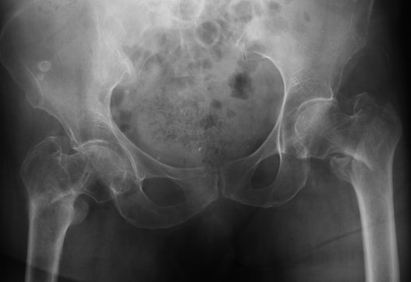
Figure 1: Intratrochanteric fracture of the left
hip.
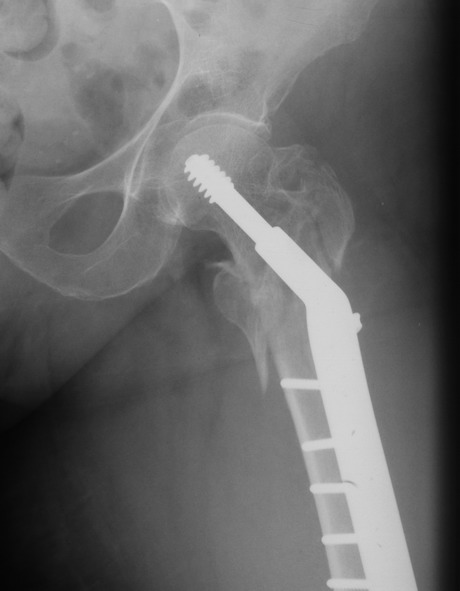
Figure 2: Dynamic hip screw fixation of the
intratrochanteric fracture.
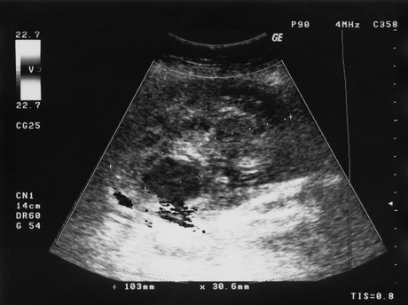
Figure 3:
Duplex ultrasound scanning identifying a 6 to 8cm haematoma
on the left femur.
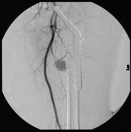
Figure 4:
CT angiogram confirmed the presence of pseudoaneurysm of medial
branch of the profunda femoral artery.
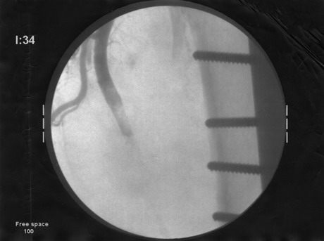
Figure 5: Embolisation of the profunda femoral
artery
As it was unclear whether the pulsatile flow was actually within
the haematoma or simply lying beneath it, coupled with evidence
of ongoing coagulation (represented by the areas of damped
flow), a conservative management approach was pursued.
Unfortunately, the patient required repeated blood transfusions
and correction of a coagulopathy over the following 10 days,
despite remaining haemodynamically unstable. We therefore
proceeded to CT angiogram, which confirmed the presence of
pseudoaneurysm of medial branch of the profunda femoral artery
(Fig. 4).
She was then successfully treated with embolisation under
fluoroscopic control in the profunda femoris artery immediately
related to the origin of the pseudoaneurysm (fig. 5). There was
no evidence of recurrence at 3-month follow-up.
Discussion :
Pseudoaneurysm of the profunda femoral artery after proximal
femoral fracture fixation is rare [2, 4, 6, 9].
Iatrogenic injury is caused by
over-penetration of the drill
bit or sharp instrument while applying dynamic
compression plate, retractor tip
pressure or by fracture edges
during manipulation [5, 10]. The diagnosis is usually
delayed because pain, haematoma and
anaemia are attributed to
post-operative complications and are non-specific [3, 4].
Clinical signs such as a tense thigh swelling, hip pain (as a
direct pressure effect), persistent or recurrent anaemia from
continued haemorrhage, neurological compromise and distal
ischaemia due to impaired blood flow or microembolisation should
suggest the possibility of false aneurysm formation. Our case
presented unusually early, as the patient was still in hospital,
recovering from her initial hip surgery.
The artery could either be lacerated or punctured by the drill
bit or sharp instrument over-penetration [5]. While applying
clamps to hold the plate against the bone the vessel might fix
against the medial wall of the femur making it more susceptible
for injury while drilling. The chances of vascular injury could
be reduced if overpenetration of the drill bit is avoided into
the medial soft tissues, using proper sized screws and careful
placement of the clamps used to hold the plate against the
femur.
There should be low threshold to diagnosis because though rare
it can be a serious complication. Diagnosis is best confirmed by
the duplex scan or a CT angiogram and endovascular embolisation
is the standard management of this condition [4].
Conclusion:
Pseudoaneurysm of the profunda femoral artery following dynamic
hip screw fixation of a femoral fracture is rare and
presentation is usually late. High index of suspicion is
required for early diagnosis. Careful use of sharp instruments,
drill bits, clamps and retractors during the operation
especially when dealing with complicated fracture pattern.
Duplex scanning often establishes the diagnosis but
arteriography may be required.
The advent of interventional radiology has allowed minimally
invasive treatment of false aneurysms and we present a case of
successful embolisation of an iatrogenic profunda femoral artery
pseudoaneurysm which is the standard management in this
condition.
Reference :
-
Barnes DI, Broude HB. False aneurysm of the profunda femoris
artery complicating fracture of the femoral shaft and treated
by transcatheter embolization. A case report. S Afr Med J.
1985; 67:824-826
-
Canbaz S, Acipayam M, Gurbuz H, Duran E. False aneurysm of
perforating branch of the profunda femoris artery after
external fixation for a complicated femur fracture. J
Cardiovasc Surg (Torino). 2002; 43:519-521
-
Chong KC, Yap EC, Lam KS, Low BY. Profunda femoris artery
pseudoaneurysm presenting with triad of thigh swelling,
bleeding and anaemia. Ann Acad Med Singapore. 2004;
33:267-269
-
Fernandez Gonzalez J, Terriza MD, Cabada T, Garcia-Araujo C.
False aneurysm of the femoral artery as a late complication of
an intertrochanteric fracture. A case report. Int Orthop.
1995; 19:187-189
-
Fordyce A. False aneurysm of the profunda femoris artery
following nail and plate fixation of an intertrochanteric
fracture. Report of a case. J Bone Joint Surg [Br].
1968; 50:141-143
-
Kleintz R, Nolte U. [Development of aneurysma spurium of the
arteria profunda femoris as a late complication of DHS
osteosynthesis]. Unfallchirurg. 1993; 96:39-40
-
Laohapoonrungsee A, Sirirungruangsarn Y, Arpornchayanon O.
Pseudoaneurysm of profunda femoris artery following internal
fixation of intertrochanteric fracture: two cases report. J
Med Assoc Thai. 2005; 88:1703-1706
-
Manner M, Rosch B, Roy K. [Vascular injuries complicating
osteosynthesis in proximal femur fractures]. Unfallchirurg.
1999; 102:227-231
-
Murphy PG, Geoghegan JG, Austin O, More-O'Ferrall R, Quinlan
WR, Keaveny TV. Pseudoaneurysm of the profunda femoris artery
due to intertrochanteric fracture of the hip. Arch Orthop
Trauma Surg. 1999; 119:117-118
-
Obry C, Mertl P, Woestelandt T, Vives P. [False aneurysm of
the profunda femoris artery after fracture of the upper end of
the femur. Apropos of a case]. Rev Chir Orthop Reparatrice
Appar Mot. 1988; 74:585-587
|

