|
Abstract:
Introduction: intra articular fractures of distal humerus
continue to be a treatment challenge. Advances in the surgical
techniques and implants have led to progressive improvement in
the outcome of these difficult fractures.
Material and Method: 164 patients with intra articular
fractures of distal humerus were treated in the Department of
Orthopedics, Government Medical College Srinagar by open
reduction and internal fixation, using trans olecranon. Patients
were followed for a minimum period of six months, maximum follow
up was 48 months. Results were assessed using scoring system of
Caja C.L and Morrani A et. al.
Result: 68 patients (42%) patients were graded as
excellent (90 to 100 points), 74 patients (45%) as good (75 to
85 points), 17 (10%) as fair (50 to 65 points) and 5 patients
(3%) as poor (less than 50 points). Level of activity was higher
in higher range of motion subgroup. Severity of fracture
affected the radiological, functional and total score. Patients
with higher radiological scores had higher range of motion and
higher activity level of activity.
Conclusion: Thorough evaluation of fracture anatomy,
meticulous surgical technique, stable fracture fixation and
early range of motion are the corn stones to restore the pre
fracture function of injured elbow.
J.Orthopaedics 2008;5(4)e9
Keywords:
Intraarticular fractures; distal humerus ; outcome.
Introduction:
The intra
articular fractures of distal end of humerus constitute about 2%
of all fractures 1. These fractures are a treatment challenge,
to the point of being intimidating and frustrating to the
operating surgeon 2,3. When these fractures extend into the
elbow joint, there is significant risk of residual pain and
functional impairment 4,5. The recommendations for the treatment
range from essentially no treatment to open reduction and
extensive internal fixation 6,7. Conservative treatment of intra
articular fractures of distal humerus usually results in loss of
elbow motion and permanent disability4,7 With the improvement
in surgical skills and implants, the outcome of these fractures
continues to improve 7.
The lack of a widely accepted scoring system makes study of
these difficult fractures even more difficult 8. A large number
of scoring systems have been proposed for the post operative
evaluation of these fractures 1,8,10,11,12,13, 14, but only a
few have used clinical and radiological parameters.6,9,10,14.
The aim of present study was to evaluate the outcome of intra
articular fractures of distal end of humerus treated by open
reduction internal fixation using trans olecranon approach and
assessed by scoring system of Caja CL and Moorani A et. al 8.
Material and Methods :
From June 2002 to October 2007, 164 patients with intra
articular fractures of distal humerus were treated in the
Department of Orthopedics, Government Medical College Srinagar
University of Kashmir, by open reduction and internal fixation,
using trans olecranon approach. There were 69 (42%) male and 95
(58%) female patients; mean age was 53 years, ranging from 14 to
90 years. Mode of injury was falls in 96 (58.5%), Road traffic
accidents in 41 (25%) and direct hit in 27 (16.5%) patients. The
fractures were classified as per AO classification into C1, C2
and C3 types. There were 72 (44%) type C1, type 60 (36.5%)C2 and
32(19.5%) type C3 fractures. 96(58.5%) fractures affected right
side and 68(41.5%) affected left side and 18(11%) fractures were
type 1 compound.
All
patients were operated within 5 days of admission using AO
technique, exposing the fracture by a dorsal skin incision and
olecranon osteotomy. In all cases the fracture was stabilized
with two plates and an intercondylar screw or a plate and a
screw in addition to the intercondylar screw. All osteotomies
were stabilized with a 6.5 or a 4.5 mm cacellous screw
reinforced with a dorsal ulnar tension band wire. Post
operatively elbow was immobilized in a crammer wire splint.
Range of motion exercises were started from the first post
operative day. The splint was removed for the day and was
re-applied at night, till wound healed and sutures were removed,
when splintage was discarded. Patients were followed weekly for
one month, bi-weekly for 3 months, then monthly for a maximum
period of 48 months (average 28 months). Postoperative
radiographs were compared and assessed for adequacy and quality
of surgical reduction. Fig 1 & 2
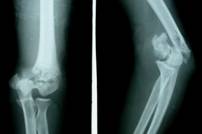
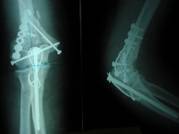
Fig.1
Type C2 Fracture
Fig.1A:
Pre operative Radiograph Fig 1B:Immediate
post operative
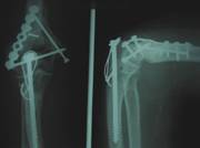
Fig
1 C:After 6 months
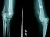
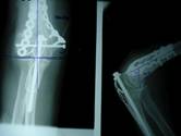
Fig .
2. Type C1Fracture
Pre operative
Radiograph
Immediate post operative
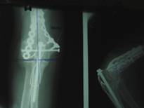
After 6 months
The parameters noted included dimensions of any articular
surface step, articular surface diastases, narrowing of distal
humeral articular surface, malalignment of AP carrying angle and
trochlea-capitellum angle, any Para articular calcification,
loosening of implant and progression of union. Range of motion,
functional status of patient, pain and complications if any were
noted. Final assessment was done at the end of 6 months using
scoring system of C L Caja and Moorani A. et al. 1994 8 It is a
100 Point scoring system and considers four parameters: pain (40
points), Range of motion (30 points), level of activity compared
to activity prior to injury (10 points) and radiological quality
of surgical reduction (20 points).
Results :
Average healing time of fractures and osteotomies was 14 weeks
(Range 9 to 20 weeks). There were two non unions at
supracondylar region which needed a secondary procedure of bone
grafting and DCP fixation. Both subsequently united and were
graded as good results. In five olecranon osteotomies union was
delayed up to 20 weeks, all of which subsequently healed without
any secondary intervention. Pain was seen in 42 patients, 17 had
pain because of prominent hardware and bursa over olecranon
screw, 23 had occasional activity related pain and 2 patients
had pain with activities of daily living. Maximum range of
motion was gained in 12 weeks, average range of motion was 100o
(range 900 to 1300). 67 (41%) patients had full range of motion,
83 patients (50%) had range of motion more than functional range
of Morrey15, 14 (9%) patients had range of motion less than
functional range.
126 (77%) patients had activity level as prior to injury; it was
diminished in 30 (18%) and restricted in 8(5%).
There were two ulnar nerve palsies, one because one backed out
screw was pressing upon the nerve, which resolved once backed
out screw was removed. In other patient ulnar nerve palsy
improved only after anterior transposition after 12 weeks of
surgery. Superficial wound infection was seen in 18 patients.
There was no deep infection.
68 patients (42%) patients were graded as excellent (90 to 100
points), 74 patients (45%) were graded as good (75 to 85
points), 17 (10%) as fair (50 to 65 points) and 5 patients (3%)
as poor (less than 50 points). Level of activity was higher in
higher range of motion subgroup. Severity of fracture affected
the radiological, functional and total score. Patients with
higher radiological scores had higher range of motion and higher
activity level. Minor complications occurred in some patients.
The radiological criteria which were difficult to maintain ,
were articular surface step more than 1 mm in 37 (38%)
fractures, anterior trochlea-capitellum angle, malalignment of
more than 100 was seen in 34 (35%) cases. Para articular
calcification of more than 10mm developed in 29 (30%) cases,
articular surface diastases of more than 1mm and malalignment of
AP carrying angle of more than 100 was observed in 6 (6%)and
11(11.5%) cases respectively. (table 1)
Table:1
|
Parameter
|
No. of patients ( % ) |
|
A. Pain |
|
|
No Pain |
104 (67%) |
|
Occasional pain |
23(30%) |
|
Activity related mild pain |
19 (3%) |
|
B. Range of motion (ROM) |
|
|
Full ROM |
67 (41%) |
|
ROM more than functional range |
83 (50%) |
|
ROM less than functional range |
14 (9%) |
|
C. Activity Level |
|
|
As prior to trauma |
126 (90%) |
|
Diminished |
30 (7%) |
|
Interrupted |
8 (3%) |
|
D. Radiological quality of surgical reduction. |
|
|
Articular surface step more than 1 mm |
38 (24%) |
|
Articular surface diastases more than 1mm |
11 (7%) |
|
AP carrying angle malalignment less than 10º |
6 (4%) |
|
Heterotrophic ossification less than 10 mm |
29(18%) |
|
anterior capitellum- trochlea angulation malalignment more
than 10º |
34(20%) |
|
E. Complications |
|
|
Superficial wound infection |
8 (5%) |
|
Ulnar nerve palsy |
2 (1%) |
|
Prominent olecranon screw |
23 (14%) |
|
Painful Bursa over screw head |
17 (6%) |
|
Secondary procedure for removal of symptomatic osteotomy
fixation |
29 (18%) |
|
Delayed union |
5 (3%) |
|
Non union |
2 (1%) |
|
(ROM= Range of motion) |
|
Severity of fracture affected radiological, functional and total
score. Patients with higher radiological score had higher
functional outcome.
Discussion :
The intra-articular fractures of distal humerus are difficult to
treat because of the nature of injury and intricate anatomy of
the region 1, 14. The recommendations for treatment range widely
from essentially no treatment to open reduction and extensive
internal fixation 11,12.The aim of operative treatment of intra-articular
fractures of distal humeral is anatomic reduction, rigid
fixation to allow early range of motion and finally to restore
the pre fracture function5,13. The quality of elbow function,
after fracture of distal humerus has been related to the degree
to which to which normal anatomic relations are restored
1,10,12,14. Elbow mobility is hindered by loss of normal
anterior tilt of distal humeral articular surface, narrowing or
distraction of distal articular surface or by obstruction of
coronoid and olecranon fossae. Pain has been related to failure
of fracture to unite, restricted motion, ulno humeral arthrosis
or instability and compression of ulnar nerve. 2,9
The anatomic reduction of articular fragments is made difficult
by poor visualization because of extensor mechanism and intact
olecranon process which is hocked over the trochlea. Direct
visualization of fracture is enhanced by mobilizing extensor
mechanism which is further enhanced by osteomatising the
olecranon process.1,5,6,9
The studies of outcome of these difficult fractures are made
even more difficult because of relative rarity; substantial
variability among different case series in terms of type of
fracture included, operative techniques and type of implants
used and method of rating results. Lack of a universally
accepted scoring system further compounds the problem
1,7,12,13,14,. Large number of scoring systems have been
proposed by numerous authors based either on the post operative
range of motion of the elbow 1112,13 or on the postoperative
range of motion, pain and disability 1,8,,13. Few authors
considered the quality of the surgical reduction as one of the
criteria in evaluation of results of these difficult fractures
6,,11,14 however there was no attempt to quantify them. Caja CL
and Moorani A developed a comprehensive 100 point scoring system
with an attempt to quantitate the quality of the surgical
reduction and the functional outcome of the patients. This
scoring system considers four parameters: pain 40 points, range
of motion 30 points, radiological quality of surgical reduction
20 points and post operative activity level 10 points. The aim
of present study was to assess outcome of these fractures using
the evaluation criteria of Caja CL and Moorani A. 8
Reference :
-
Jupiter JB, Neff U, Holzach P, et al. Intercondylar
fractures of distal end of humerus. An operative approach. J
Bone Joint Surg Am. 1985; 67A: 226239.
-
Driscoll SW. Triceps reflecting anconeus pedicle approach
for distal humeral fractures and non unions. Orthop clin North
Am. 2000; 31: 91.
-
Aitken GK, Roraback C.H: distal humeral fractures in the
adult. Clinc Orthop. 1988; 207: 191.
-
Bickel W. H and Perry R.E. Comminuted fractures of distal
humerus. J.A.M.A.1963; 184: 553.
-
Helfert DL. Bicondylar intraarticular fractures of distal
humerus in adults: their assessment, classification and
operative management. Adv Orthop surg.1985; 8:223-235.
-
Holdsworth BJ, Mossad MM. Fractures of adult distal
humerus. Elbow function after internal fixation. J Bone Joint
Surg Br. 1990; 72B:362-365.
-
Dowden JH. Principle of early active movement in treating
fractures of upper extremity. Clin Orthop. 1981; 148: 4.
-
Caja VL, Moroni A, Vendemia V. et al. surgical treatment
of bicondylar fractures of distal humerus. Injury 1994; 25:
433-438.
-
Cassebaum WH. Open reduction of T & Y fractures of lower
end of humerus. J Trauma. 1969; 9:915-925.
-
Letch R, Schmit-Neuberg P. Intra articular fractures of
distal humerus: surgical treatment and result. Clin orthop.
1989; 241:238
-
Gabel GT, Hanson G, Bennet JB, et al. Intraarticular
fractures of distal humerus in the adult. Clin Orthop.
1987;216:99.
-
Horne G. Supracondylar fractures of humerus in adults. J
Trauma .1980;20(11):71.
-
Martin J, Marsh JH, Nepola JV. Radiographic fracture
assessment: which ones we can reliably make? J Orthop Trauma.
2000; 14:379-385.
-
Ring D, Jupiter JB. Fractures of the distal humerus.
Orthop clinic North Am. 2000; 31: 103.
-
Morrey BF, Askew LJ, et al. A biomechanical study of normal
functional elbow. J Bone Joint surg Am. 1981; 63A: 872-876.
|

