|
Abstract:
There is a possible
relationship between joint replacements and malignancy. We
review the current state of metal-on-metal hip implants, the
process of metal ion release from the implants, the effect of
released metal ions in vivo, the association between
neoplasm and metal debris and ions, and the six documented cases
of neoplasm around a metal-on-metal total hip replacement. We
find no link between released metal ions and the occurrence of
neoplasm.
J.Orthopaedics 2008;5(3)e9
Keywords:
metal-on-metal hip implants, metal ion
release, metal debris, total hip arthroplasty, total hip
replacement, neoplasm, malignancy, cancer.
Introduction:
Total hip arthroplasty (THA) has evolved into one of the most
common and cost-effective orthopedic reconstructive procedures
with a high long-term success rate. However, due to the dynamic
nature of the hip implants and the direct contact with
biological tissue, particulate wear and corrosion debris are
liberated into the joint space and surrounding tissues 1, 2. The
subsequent biological response poses a problem to the durability
and long-term efficacy of the hip implants. Metal ions that are
released, such as cobalt and chromium, are of importance because
of their carcinogenic potential3, 4. Of particular interest are
hip implants with metal-on-metal bearings (MOM). There have been
several cases of malignant tumors that have developed at the
site of such implants. The purpose of this review is to examine
if there is any connection between neoplasia and metal ion
debris in THA. The following review is organized into six
sections: (1) current state of MOM THAs, (2) metal bearing wear
and ion release, (3) biology of ions, (4) neoplasia associated
with metal debris and ions, (5) reported cases of neoplasia
around THA (6) conclusion.
Current State of MOM THA
Metal-on-metal (MOM) bearing coupling was popular in the 1960s
and 1970s, but was abandoned by US surgeons in favor of
metal-on-polyethylene bearings due to concerns over biological
reactions and reported high loosening rates. One of the
limitations of polyethylene is wear debris generation at the
bearing surface. This led to the development of alternative
bearing surfaces including the reintroduction of MOM bearings in
the late 1980s5. These newer designs with MOM bearings have
been shown to be clinically efficacious and more durable than
earlier designs. Dorr et al.6 reported mechanical failure rates
(combined rates of revision and loosening) in 56 patients with
the Metasul (Zimmer, Warsaw, IN) design from 1991 to 1994. There
were 49 patients with primary osteoarthritis, 3 with congenital
dysplasia, and 2 each with posttraumatic arthritis and femoral
head osteonecrosis. All patients had well functioning hips at an
average of 5.2 years of follow-up.
Wear debris and metal ion release are clinical concerns. Jacobs
et al.7 reported 3-fold serum concentration increases in
titanium and 5-fold increases in chromium at thirty-six months
following surgery in 55 patients with MOM THAs compared to
control patients without implants. Sauve et al.8 reported 5-fold
and 3-fold serum concentration increases for cobalt and
chromium, respectively in 310 patients with Ring MOM THAs as
long as 30 years after the index surgery. It is of interest to
note that in 3 of the patients who underwent revision for
aseptic loosening, serum metal concentrations were within normal
range.
Meat bearing wear is influenced by several factors: carbon
content, manufacturing process, and diametral clearance. Rieker
et al.9 reported on analysis of a large collection of retrieved
MOM components. These included 608 components from 337
revisions. 172 of these were analyzed for clearance (surface
geometry match between the head and the liner). These components
were in-vivo from one month to 12 years. They reported several
important findings: 1) the mean wear rate in the first year was
high (27.8 um/year), 2) the mean wear rate following the second
year was low (6.2 um/year), 3) regression analysis showed
clearance to be the most important variable correlated with the
linear wear rate (p=0.0005). Current laboratory and clinical
retrieval data on MOM couplings support the idea that superior
wear characteristics are associated with high carbon content,
wrought manufacturing, large diameter, and low clearance.
Metal Bearing Wear and Ion Release
Metal ions release can occur by two proposed mechanisms: 1) wear
as a function of adhesion, abrasion, or fatigue of the materials
resulting in particulate debris, and 2) corrosion of the metal
bearing wear resulting in the release of metal ions. Metallic
implants have the ability to achieve passivity in-vivo, in which
active and passive surfaces between metallic biomaterials and
electrolytes exist in contact simultaneously. The protective
surface oxide layer on the implants is in a continuous process
of partial dissolution and reprecipitation10. Dissolution is
favored by contact with proteins and amino acids leading to
corrosion. In addition, macrophages that ingest released
particles are stimulated by inflammatory mediators such as TNF-α
to release reactive oxygen species such as superoxide (O2-) and
hydrogen peroxide (H2O2). These products can be metabolized into
products such as hypoclorous acid, capable of damaging
extracellular matrix components and increasing degradation by
proteases11.
High serum and urine metal ion concentrations have been
associated with both first and second generation MOM bearings.
Jacobs et al.12 found that serum chromium concentration was
9-fold higher and cobalt concentration 3-fold higher in patients
with McKee-Farrar THAs than the control patients without
implants. Second generation implants showed similar increases in
metal ions. Savarino et al.13 reported 4-to-5-fold higher cobalt
concentrations and 7-fold higher chromium concentrations in
patients with the Metasul THAs than in control patients without
implants.
There is concern over metal ion release in the growing
population of young and active patients that receive THAs.
Heisel et al.14 compared cobalt and chromium levels in patients
with MOM THAs as a function of varying levels of activity. They
found a 2.7% and 3.0% serum concentration increase in cobalt and
chromium, respectively when the patients increased their mean
activity by 28% as measured by a 2-dimensional accelerometer
worn on the ankle. During treadmill running, mean activity
increased by 1621% while serum cobalt and chromium
concentrations increased by 3.0% and 0.8%. Based on the accuracy
of the tests and the small relative size of serum metal ion
changes, the data did not demonstrate statistically significant
correlation between activity level and serum metal ions.
Release of metal ions from MOM THAs has clinical significance
for child-bearing females. Ziaee et al.15 collected blood from
the mother and umbilical cord after five deliveries, four of
which involved MOM THAs. The chromium and cobalt levels were an
average of 50% lower in the cord than in maternal blood and
there was a strong correlation between levels of trace elements
in the cord and maternal blood.
Biology of Ions
Metal ion toxicity can occur if the ion binds with a biomolecule.
Titanium ions are very active and immediately bind to water or
inorganic anions within the tissue. On the other hand, inactive
ions such as copper and nickel remain as ions for long durations
and have a greater chance of combining with biomolecules to
express toxicity10. Trace elements that show similar
high-reactive ion binding qualities to titanium, such as
zirconium, niobium, and tantalum, hold promise as alloys that
could decrease bonds between metal ions and biomolecules.
Released ions would instead bind harmlessly to water and
inorganic anions in the tissue.
Metal ion toxicity can lead to local tissue inflammation,
fibrosis, and necrosis. Mathiesen et al.16 reported extensive
periprosthetic tissue necrosis due to metal ion toxicity in 4 of
9 patients with MOM THAs. Metal particles can also be found
beyond the periprosthetic tissue to regional lymph nodes, liver,
and spleen12, 17, 18. These particles can interfere with
osteoblasts and osteoclasts, inducing bone resorption and cause
toxicity to macrophages and fibroblasts19, 20.
Several recent studies have documented evidence of
hypersensitivity to metal wear particles in patients with
painful and failed MOM THAs. Park et al.21 reported a
significantly higher prevalence of hypersensitivity to cobalt
(p=0.03) in patients with early osteolysis when compared to
control patients. The cohort consisted of 169 THAs with
second-generation MOM bearings, with ten reported cases of
osteolysis. Histiologic analysis of periprosthetic tissue from
two patients with osteolysis revealed a perivascular
accumulation of CD3-positive T-cells and CD68-positive
macrophages. Immunohistochemical analysis demonstrated the
presence of bone-resorbing cytokines such as IL-1β and TNF-α.
These findings suggest that delayed hypersensitivity in MOM THAs
may cause early osteolysis. Milosev et al.22 reported similar
evidence of osteolysis from hypersensitivity to metal wear
particles. He analyzed a cohort of 591 patients with MOM THAs at
a mean of seven years postoperatively and showed a survival rate
91% at ten years (95% confidence interval 0.88 to 0.95). The
major cause of failure was aseptic loosening, which occurred in
25 patients. Histological analysis of 17 of the revision THAs
showed a hypersensitive reaction in 13 patients (2%), with
perivascular infiltration of lymphocytes and aseptic
inflammation.
Neoplasia Associated with Metallic Debris and Ions
Metal ions have been linked to increased cancer rates. Visuri et
al.23 reported a standardized incidence ratio (rate of
occurrence in the cohort compared to the general population) of
3.0 (95% confidence interval 1.1-6.6) for hematopoietic cancers
in a cohort of 433 patients with McKee-Farrar MOM THAs. The
standardized incidence rate of leukemia was also elevated at 3.2
(95% confidence interval 1.0-7.4).
Current MOM THAs are made of alloy biomaterials consisting of
cobalt, chromium, titanium, aluminum, vanadium, and nickel. The
carcinogenic potential of cobalt and nickel have been reported
in animals. Intramuscular or intrathoracic injections of cobalt
metal powder produced fibrosarcomas and rhabdomyosarcomas at the
injection sites in mice while intratracheal instillation of
nickel metal powder in rats resulted in significant numbers of
squamos-cell carcinomas and adenocarcinomas of the lung24.
Osteosarcomas and fibrosarcomas have also been found in cats and
dogs with stainless-steel metallic composition internal fixation
devices25. However, Lewis et al.26 injected CoCrMo or TiAlV wear
debris powder into the knee joints in rats. They found no case
of neoplasia as a result of this exposure.
The exact mechanisms and association between neoplasia and
metallic trace elements remain to be defined. The association of
implant loosening and tumors around the implant site in rats
suggests that neoplasia resulted from a foreign-body reaction27.
This relationship has yet to be established in humans.
Macrophages binding to implants result in the generation of
reactive oxygen species (ROS)28, 29. ROS can cause alterations
in host DNA, most frequently causing guanine-to-thymine
transversions30. If these modifications are associated with
oncogenes or tumor suppressor genes, then metal ion release can
indirectly lead to the pathogenesis of neoplasia. Multiple
mutations in the p53 tumor suppressor gene have been reported as
guanine-to-thymine transversions31, 32 and oxidative DNA
modifications in cancer tissue from ROS have been reported33.
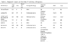
Reported cases of neoplasia
Since 1974, there have been 51 reported cases of malignant
tumors at the site of a THA in the English literature. These
include 20 cases of malignant fibrous histiocytoma, 10
osteosarcoma, 4 non-Hogkin lymphoma, 3 spindle cell sarcoma, and
2 cases each of pleoemorphic rhabdomyosarcoma, fibrosarcoma, and
leiomyosarcoma. Other individual cases were chondrosarcoma,
malignant peripheral nerve sheath tumor, synovial sarcoma,
multiple myeloma, liposarcoma, adenocarinoma and malignant
epithelioid hemangioma. The patients range in age from 39 to 88
years, with a mean age of 66 years. The mean time interval from
implantation to the diagnosis of neoplasia was 6.6 years with a
range of 0.5 to 20 years. 6 of these occurred around MOM THAs.
We will detail these 6 cases.
Penman and Ring34 reported a case of ostesarcoma in a
75-year-old woman with a cobalt-chromium MOM THA. The tumor was
discovered 5 years post-operatively on the lateral side of the
right femur. There was no history of Pagets disease or
radiotherapy. The authors concluded that metal ion release could
have been associated with the development of the tumor.
Swann35 reported a case of malignant fibrous histiocytoma in a
63 year-old man that developed 3.5 years post-operative to a MOM
THA. Ryu et al.36 reported a case of soft tissue sarcoma in a 40
year-old man 1.5 years post-operative to a MOM THA. Stephenson
et al.37 reported a case of liposarcoma in a 57 year-old man 6
years post-operative to a MOM THA. In all 3 of these cases, the
tumors were not in direct contact with the metal implants. Metal
content was only measured in the liposarcoma patient and no
increased levels were found. This evidence suggested that the
tumors were not directly caused by the carcinogenic potential of
metal ions or debris from the implants.
Rushford38 reported a case of osteosarcoma in the acetabulum 5
months post-operative to a cobalt-chromium MOM THA in a
54-year-old woman. She had previously developed squamos cell
carcinoma in her cervix which was treated successfully with
radium insertion and external irradiation. The prosthetic cup
was found to be loose and the acetabulum bone quality was
abnormal. The authors concluded, based on the greater proportion
of bone radiation absorption, that the patients previous
radiotherapy resulted in the subsequent development of
osteosarcoma.
Arden and Bywater39 reported a case of fibrosarcoma 2.5 years
post-operative to a MOM THA in a 56-year-old patient. The
sarcoma was found to arise from the arthroplasty scar and extend
down to the pseudo-capsule, but did not involve the femur or
pelvis bones.
Conclusion:
There is no
conclusive evidence to establish any direct link between metal
ion or debris with malignancy around THAs with MOM bearings. It
is however critical for these participants to be followed,
especially when MOM THAs are being done with greater frequency
and in younger patients.
Table 1.
Malignant Tumors at the Site of Total Hip Arthroplasty
(click to enlarge)
Reference :
1. Jacobs JJ, Gilbert JL, Urban RM. Corrosion of metal
orthopaedic implants. J Bone Joint Surg Am 1998;80(2):268-82.
2. Jacobs JJ, Hallab NJ, Urban RM, Wimmer MA. Wear particles. J
Bone Joint Surg Am 2006;88 Suppl 2:99-102.
3. Katz SA, Salem H. The toxicology of chromium with respect to
its chemical speciation: a review. J Appl Toxicol
1993;13(3):217-24.
4. Lison D, De Boeck M, Verougstraete V, Kirsch-Volders M.
Update on the genotoxicity and carcinogenicity of cobalt
compounds. Occup Environ Med 2001;58(10):619-25.
5. Dumbleton JH, Manley MT. Metal-on-Metal total hip
replacement: what does the literature say? J Arthroplasty
2005;20(2):174-88.
6. Dorr LD, Wan Z, Longjohn DB, Dubois B, Murken R. Total hip
arthroplasty with use of the Metasul metal-on-metal
articulation. Four to seven-year results. J Bone Joint Surg Am
2000;82(6):789-98.
7. Jacobs JJ, Skipor AK, Patterson LM, et al. Metal release in
patients who have had a primary total hip arthroplasty. A
prospective, controlled, longitudinal study. J Bone Joint Surg
Am 1998;80(10):1447-58.
8. Sauve P, Mountney J, Khan T, De Beer J, Higgins B, Grover M.
Metal ion levels after metal-on-metal Ring total hip
replacement: a 30-year follow-up study. J Bone Joint Surg Br
2007;89(5):586-90.
9. Rieker C, Koettig P, Schoen Rea. Clinical tribological
performance of 144 metal-on-metal hip articulations. In: CB R, M
W, U W, eds. Metasul: a metal-on-metal bearing. Bern
(Switzerland): Hans Huber; 1999:83.
10. Hanawa T. Metal ion release from metal implants. Material
Science and Engineering 2004;24(6-8):745-52
11. Wang ML, Hauschka PV, Tuan RS, Steinbeck MJ. Exposure to
particles stimulates superoxide production by human THP-1
macrophages and avian HD-11EM osteoclasts activated by tumor
necrosis factor-alpha and PMA. J Arthroplasty 2002;17(3):335-46.
12. Jacobs JJ, Skipor AK, Doorn PF, et al. Cobalt and chromium
concentrations in patients with metal on metal total hip
replacements. Clin Orthop Relat Res 1996(329 Suppl):S256-63.
13. Savarino L, Granchi D, Ciapetti G, et al. Ion release in
patients with metal-on-metal hip bearings in total joint
replacement: a comparison with metal-on-polyethylene bearings. J
Biomed Mater Res 2002;63(5):467-74.
14. Heisel C, Silva M, Skipor AK, Jacobs JJ, Schmalzried TP. The
relationship between activity and ions in patients with
metal-on-metal bearing hip prostheses. J Bone Joint Surg Am
2005;87(4):781-7.
15. Ziaee H, Daniel J, Datta AK, Blunt S, McMinn DJ.
Transplacental transfer of cobalt and chromium in patients with
metal-on-metal hip arthroplasty: a controlled study. J Bone
Joint Surg Br 2007;89(3):301-5.
16. Mathiesen EB, Lindgren JU, Blomgren GG, Reinholt FP.
Corrosion of modular hip prostheses. J Bone Joint Surg Br
1991;73(4):569-75.
17. Urban RM, Jacobs JJ, Tomlinson MJ, Gavrilovic J, Black J,
Peoc'h M. Dissemination of wear particles to the liver, spleen,
and abdominal lymph nodes of patients with hip or knee
replacement. J Bone Joint Surg Am 2000;82(4):457-76.
18. Case CP, Langkamer VG, James C, et al. Widespread
dissemination of metal debris from implants. J Bone Joint Surg
Br 1994;76(5):701-12.
19. Haynes DR, Rogers SD, Hay S, Pearcy MJ, Howie DW. The
differences in toxicity and release of bone-resorbing mediators
induced by titanium and cobalt-chromium-alloy wear particles. J
Bone Joint Surg Am 1993;75(6):825-34.
20. Goodman S, Aspenberg P, Song Y, et al. Tissue ingrowth and
differentiation in the bone-harvest chamber in the presence of
cobalt-chromium-alloy and high-density-polyethylene particles. J
Bone Joint Surg Am 1995;77(7):1025-35.
21. Park YS, Moon YW, Lim SJ, Yang JM, Ahn G, Choi YL. Early
osteolysis following second-generation metal-on-metal hip
replacement. J Bone Joint Surg Am 2005;87(7):1515-21.
22. Milosev I, Trebse R, Kovac S, Cor A, Pisot V. Survivorship
and retrieval analysis of Sikomet metal-on-metal total hip
replacements at a mean of seven years. J Bone Joint Surg Am
2006;88(6):1173-82.
23. Visuri TI, Pukkala E, Pulkkinen P, Paavolainen P. Cancer
incidence and causes of death among total hip replacement
patients: a review based on Nordic cohorts with a special
emphasis on metal-on-metal bearings. Proc Inst Mech Eng [H]
2006;220(2):399-407.
24. McGregor DB, Baan RA, Partensky C, Rice JM, Wilbourn JD.
Evaluation of the carcinogenic risks to humans associated with
surgical implants and other foreign bodies - a report of an IARC
Monographs Programme Meeting. International Agency for Research
on Cancer. Eur J Cancer 2000;36(3):307-13.
25. Black J. Orthopedic Biomaterials in Research and Practice.
New York: Churchill Livingstone; 1988.
26. Lewis CG, Belniak RM, Plowman MC, Hopfer SM, Knight JA,
Sunderman FW, Jr. Intraarticular carcinogenesis bioassays of
CoCrMo and TiAlV alloys in rats. J Arthroplasty
1995;10(1):75-82.
27. Bouchard PR, Black J, Albrecht BA, Kaderly RE, Galante JO,
Pauli BU. Carcinogenicity of CoCrMo (F-75) implants in the rat.
J Biomed Mater Res 1996;32(1):37-44.
28. Adams DO, Hamilton TA. The cell biology of macrophage
activation. Annu Rev Immunol 1984;2:283-318.
29. Cross AR, Jones OT. Enzymic mechanisms of superoxide
production. Biochim Biophys Acta 1991;1057(3):281-98.
30. Waris G, Ahsan H. Reactive oxygen species: role in the
development of cancer and various chronic conditions. J Carcinog
2006;5:14.
31. Hollstein M, Sidransky D, Vogelstein B, Harris CC. p53
mutations in human cancers. Science 1991;253(5015):49-53.
32. Harris CC, Hollstein M. Clinical implications of the p53
tumor-suppressor gene. N Engl J Med 1993;329(18):1318-27.
33. Poulsen HE, Prieme H, Loft S. Role of oxidative DNA damage
in cancer initiation and promotion. Eur J Cancer Prev
1998;7(1):9-16.
34. Penman HG, Ring PA. Osteosarcoma in association with total
hip replacement. J Bone Joint Surg Br 1984;66(5):632-4.
35. Swann M. Malignant soft-tissue tumour at the site of a total
hip replacement. J Bone Joint Surg Br 1984;66(5):629-31.
36. Ryu RK, Bovill EG, Jr., Skinner HB, Murray WR. Soft tissue
sarcoma associated with aluminum oxide ceramic total hip
arthroplasty. A case report. Clin Orthop Relat Res
1987(216):207-12.
37. Stephensen SL, Schwarz Lausten G, Thomsen HS, Bjerregaard B.
Liposarcoma in association with total hip replacement. Int
Orthop 1999;23(3):187-9.
38. Rushforth GF. Osteosarcoma of the pelvis following
radiotherapy for carcinoma of the cervix. Br J Radiol
1974;47(554):149-52.
39. Arden G, Bywater E. Tissue Reaction. In: Arden G, Ansell B,
eds. Surgical management of juvenile chronic polyarthritis.
London: Academic Press; 1978:269.
40. Aboulafia AJ, Littelton K, Shmookler B, Malawer MM.
Malignant fibrous histiocytoma at the site of hip replacement in
association with chronic infection. Orthop Rev
1994;23(5):427-32.
41. Adams JE, Jaffe KA, Lemons JE, Siegal GP. Prosthetic implant
associated sarcomas: a case report emphasizing surface
evaluation and spectroscopic trace metal analysis. Ann Diagn
Pathol 2003;7(1):35-46.
42. Bago-Granell J, Aguirre-Canyadell M, Nardi J, Tallada N.
Malignant fibrous histiocytoma of bone at the site of a total
hip arthroplasty. A case report. J Bone Joint Surg Br
1984;66(1):38-40.
43. Bell RS, Hopyan S, Davis AM, Kandel R, Gross AE. Sarcoma of
bone-cement membrane: a case report and review of the
literature. Can J Surg 1997;40(1):51-5.
44. Bower T. Osteosarcoma at the site of total hip replacement.
Trans Society Biomaterials 1987;10(36)
45. Brien WW, Salvati EA, Healey JH, Bansal M, Ghelman B, Betts
F. Osteogenic sarcoma arising in the area of a total hip
replacement. A case report. J Bone Joint Surg Am
1990;72(7):1097-9.
46. Cole BJ, Schultz E, Smilari TF, Hajdu SI, Krauss ES.
Malignant fibrous histiocytoma at the site of a total hip
replacement: review of the literature and case report. Skeletal
Radiol 1997;26(9):559-63.
47. Ganapathi M, Lake DN, Griffiths AP. Periprosthetic
high-grade B-cell lymphoma complicating an infected revision
total hip arthroplasty. J Arthroplasty 2001;16(2):229-32.
48. Goodfellow J. Malignancy and joint replacement. J Bone Joint
Surg Br 1992;74(5):645.
49. Haag M, Adler CP. Malignant fibrous histiocytoma in
association with hip replacement. J Bone Joint Surg Br
1989;71(4):701.
50. Harris WR. Chondrosarcoma complicating total hip
arthroplasty in Maffucci's syndrome. Clin Orthop Relat Res
1990(260):212-4.
51. Ito H, Shimizu A. Malignant lymphoma at the site of a total
hip replacement. Orthopedics 1999;22(1):82-4.
52. Jacobs JJ, Rosenbaum DH, Hay RM, Gitelis S, Black J. Early
sarcomatous degeneration near a cementless hip replacement. A
case report and review. J Bone Joint Surg Br 1992;74(5):740-4
53. Keel SB, Jaffe KA, Petur Nielsen G, Rosenberg AE.
Orthopaedic implant-related sarcoma: a study of twelve cases.
Mod Pathol 2001;14(10):969-77.
54. Lamovec J, Zidar A, Cucek-Plenicar M. Synovial sarcoma
associated with total hip replacement. A case report. J Bone
Joint Surg Am 1988;70(10):1558-60.
55. Langkamer VG, Case CP, Collins C, et al. Tumors around
implants. J Arthroplasty 1997;12(7):812-8.
56. Martin A, Bauer TW, Manley MT, Marks KE. Osteosarcoma at the
site of total hip replacement. A case report. J Bone Joint Surg
Am 1988;70(10):1561-7.
57. Mathiesen EB, Ahlbom A, Bermann G, Lindgren JU. Total hip
replacement and cancer. A cohort study. J Bone Joint Surg Br
1995;77(3):345-50.
58. Nelson JP, Phillips PH. Malignant fibrous histiocytoma
associated with total hip replacement. A case report. Orthop Rev
1990;19(12):1078-80.
59. Prasad PS, Latham JB, Tucker JK, Ball RY. Disseminated
osteosarcoma arising in the pelvis after total hip arthroplasty.
J Arthroplasty 2002;17(3):373-8.
60. Rana B, Shetty S, Grigoris P, Reid R. Sarcoma arising
adjacent to a total hip arthroplasty. Scott Med J
2001;46(1):17-9.
61. Radhi JM, Ibrahiem K, al-Tweigeri T. Soft tissue malignant
lymphoma at sites of previous surgery. J Clin Pathol
1998;51(8):629-32.
62. Rock M. Toxicity oncogenesis, case reports. In: Morrey B,
ed. Biological, material and mechanical considerations of joint
replacement. New York: Raven Press Ltd.; 1993:339.
63. Shaw JA. Multiple myeloma. A differential consideration for
osteolysis surrounding total hip arthroplasties. J Arthroplasty
1995;10(3):397-400.
64. Schmidt AH, Walker G, Kyle RF, Thompson RC, Jr.
Periprosthetic metastatic carcinoma. Pitfalls in the management
of two cases initially diagnosed as osteolysis. J Arthroplasty
1996;11(5):613-9.
65. Schuh A, Zeiler G, Holzwarth U, Aigner T. Malignant fibrous
histiocytoma at the site of a total hip arthroplasty. Clin
Orthop Relat Res 2004(425):218-22.
66. Solomon MI, Sekel R. Total hip arthroplasty complicated by a
malignant fibrous histiocytoma. A case report. J Arthroplasty
1992;7(4):549-50.
67. Syed AA, Agarwal M, Fenelon G, Toner M. Osseous malignant
non-Hodgkin's B-cell lymphoma associated with total hip
replacement. Leuk Lymphoma 2002;43(11):2213-6.
68. Tait NP, Hacking PM, Malcolm AJ. Malignant fibrous
histiocytoma occurring at the site of a previous total hip
replacement. Br J Radiol 1988;61(721):73-6.
69. Troop JK, Mallory TH, Fisher DA, Vaughn BK. Malignant
fibrous histiocytoma after total hip arthroplasty. A case
report. Clin Orthop Relat Res 1990(253):297-300.
70. van der List JJ, van Horn JR, Slooff TJ, ten Cate LN.
Malignant epithelioid hemangioendothelioma at the site of a hip
prosthesis. Acta Orthop Scand 1988;59(3):328-30.
|

