|
Gella
S *, Cooper AP **, Tulwa N #
*
Clinical
research fellow,
** Senior
House officer
#Consultant Orthopaedic Surgeon
Department of Orthopaedics,Pinderfields
General
Hospital
,
Wakefield
,
UK
Address for Correspondence:
MR
S Gella
6 Riverdale crescent
Wakefield
,
WF3 4JZ
United Kingdom
Tel: 01924 360735
Email: mrgella@hotmail.co.uk
|
|
Abstract:
We
report a rare case of a 12 year old boy who sustained a
traumatic pseudodislocation of the lateral epiphysis of the
clavicle mimicking type IV acromioclavicular dislocation. He was
managed operatively with internal fixation. Pseudodislocation and complete disruption of the
periosteal sleeve are extremely rare features and make this
unusual case very interesting.
J.Orthopaedics 2008;5(1)e13
Keywords:
Carpal tunnel syndrome, Median nerve, wrist sonography, carpal
tunnel diameter.
Introduction:
True
dislocations of the acromioclavicular joint in children are very
rare. Pseudodislocation
(distal epiphyseal-metaphyseal separation) mimics the adult form
of acromioclavicular dislocation in its behaviour1.
In particular, pseudodislocations mimicking Type IV variety
acromioclavicular joint dislocations are very rare and present
an interesting challenge for the surgeon. We report a case of distal epiphyseal injury of the
clavicle, mimicking the Type IV acromioclavicular joint
dislocation, treated with open reduction and stabilisation with
a Kirschener wire. In
the literature, only one similar case of pseudodislocation has
been reported, however in contrast to the case we present here,
the previous example was not severe enough to require open
reduction2. Cases
such as this are seldom seen and as such their management raises
interesting issues, which we highlight here.
Case
Report :
A
12 year old boy attended the accident and emergency department
after he injured his left non-dominant shoulder region when he
fell off his bicycle whilst attempting a stunt. He complained of immediate pain in his shoulder and a
reduced range of movement, limited mainly by discomfort. On examination, the left shoulder was swollen with a loss
of the clavicular prominence on the lateral aspect. The acromion process was readily palpable with marked
tenderness. A bony
prominence could be felt posteriorly within the trapezius
muscle. The skin was
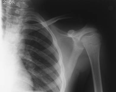
intact and he had no neurovascular deficit. Anteroposterior and axial X-ray radiographs (Figure 1)
showed left distal epiphyseal separation of the clavicle
mimicking a posteriorly displaced type IV acromioclavicular
dislocation3. The patient was treated with a broad
arm sling and admitted for further management.

Fig
1 A
Fig
1 B
Figure
1. Initial axial and antero-posterior radiographs showing a
grossly displaced clavicle
Closed
reduction was attempted under general anaesthesia but was
unsuccessful. Open reduction of the fracture through a bra-strap
incision revealed an intact acromioclavicular joint with
epiphyseal-metaphyseal separation at the lateral end of the
clavicle, corresponding to a Salter-Harris Type I injury.
The distal aspect of the clavicle was found penetrating
the trapezius muscle. The
displacement of the clavicle had resulted in extensive immediate
soft tissue damage causing complete disruption of the
periosteum. As a
result the reduction was unstable and it was not possible to
repair the periosteum as a sleeve around the clavicle. We
therefore used a 1.6mm Kirschener wire to fix the clavicle to
epiphysis (Fig 2).
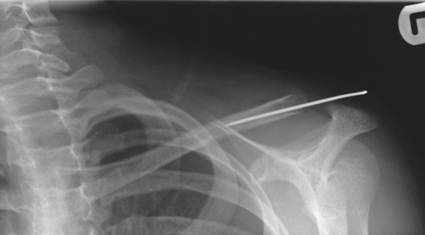
Figure
2. Anteroposterior radiograph showing K wire fixation one
week post procedure
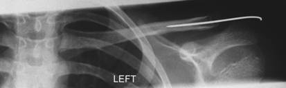
Figure
3. Anteroposterior radiograph showing bone healing and good
position at 6 weeks
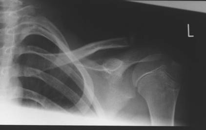
Figure
4. Anteroposterior radiograph at eight weeks post fixation
revealing bone healing in position
He
was discharged home in an arm brace and was reviewed at one and
six weeks, with check X rays revealing no loss of position and
good bone healing by six weeks (Fig. 3). At six weeks the K wire was removed and the patient was
allowed to mobilise his arm.
At
further follow-up, he was able to comfortably perform his
activities of daily living, had good overhead range of movement
and no bony deformity of the clavicle. X ray revealed that the clavicle had healed in correct
alignment with no shortening (Fig. 4).
Discussion :
Clavicular
fractures are the most common fractures to occur in childhood. Of these they most frequently occur in the middle
one-third of the shaft. Distal
epiphyseal separations are relatively uncommon injuries4. Dameron and Rockwood classified these in a scheme
mimicking the adult acromioclavicular joint dislocation3. Injuries mimicking Type IV dislocations need special
consideration as they can be easily mistaken for Type II or Type
III injuries on anteroposterior X-rays. The significance of this is that the treatment of these
varies significantly as does the potential for neurovascular
injury. Type IV injuries which protrude deep into the muscle
fibres are far less likely to unite properly without
intervention and increase the risk of injury to important
structures5. Furthermore,
because of the presence of coracoclavicular ligamentous
attachment, the periosteum can be completely peeled off when the
clavicle becomes displaced. This combination of complete periosteal separation with
the lateral end penetrating the trapezius muscle makes closed
reduction an impossible task for severe degrees of Type IV
injuries as seen in this case. In the literature there is only one other case report of
Type IV pseudodislocation of the acromioclavicular joint
described which was not severe enough to necessitate open
reduction2. Rockwood and Green describe internal fixation by means of
repair to the periosteal sleeve for fractures of the distal
clavicle3. Richard
and Howard describe a case where they were able to successfully
achieve closed reduction of a fractured clavicle, which had
buttonholed through the periosteal sleeve and penetrated the
trapezius muscle2. However in our case, the periosteum was too badly
disrupted to allow repair. This
is further substantiated by the fact that the follow-up X-ray
shows a lack of periosteal reaction around the clavicle (See
Fig. 4).
Epiphyseal
fractures such as this are of particular significance since the
clavicle has two epiphyseal areas which ossify relatively late
on in skeletal maturation. The
lateral plate fuses at age 19 and the medial at 25 6,
consequently an epiphyseal injury in a 12 year old boy leaves
him at risk of growth arrest, bony deformity and possible
limitation of function of the affected arm if not treated
appropriately.
Reference :
-
Black GB, McPherson JAM, Reed MH. Traumatic pseudodislocation of the acromioclavicular joint in children; a fifteen year review. Am J Sports Med 1991;19:644-646
-
Richards DP, Howard A. Distal clavicle fracture mimicking type IV acromioclavicular joint injury in the skeletally immature adult. Clinical J Sport Med 2001; 11:57-59
-
Sanders JO, Rockwood CA, Curtis RJ. Fractures and dislocations of the humeral shaft and shoulder. In: Rockwood CA, Wilkins KE, Beaty JH, eds. Fractures in Children. Philadelphia: J. B. Lippincott, 1991:970-977
-
Havaranek P. Injuries of distal clavicular physis in children. J Pediatr Orthop 1989:213-215
-
Dartoy C, Fennoll B, Hra D, Le-Nen D, Dubrana F, Jehannin B. Epiphyseal fracture-avulsion of the distal extremity of the clavicle. Apropos of a case Ann Radiol 1993:36:125-128
-
Gray H, The Clavicle In: Anatomy Of the Human Body 20th ed, Philadelphia: Lea & Febiger, 2000:1396
|

