|
Abstract:
Pectoralis major
tendon ruptures are uncommon injuries and most physicians have
little experience with the surgical treatment of this injury.
The purpose of this article is to describe a new method of
fixation of the ruptured tendon that facilitates the surgical
procedure without altering the postoperative care,
rehabilitation, and complication rate.
Keywords:
Pectoralis
Major, Rupture, Absorbable Anchor Sutures.
J.Orthopaedics 2007;4(4)e9
index.htm
Introduction:
Pectoralis major
tendon ruptures is a relatively uncommon injury, with only about
200 reported cases in the literature. The injury was originally
described by Patissier in 1822.1 Recently, the
reported incidence has been increasing which may be related to
the increasingly active population.
The injury has
been commonly associated with athletic competition and
weight-lifting. The mechanism is usually indirect, and
classically described as occurring when the muscle transitions
from an eccentric load to a concentric contraction. Blunt force
and traction injuries have also been described. 2, 3
The past treatment
of pectoralis major ruptures have been controversial, however
recently most authors advocate early repair.3, 4
Several studies have demonstrated significant strength deficits
with persistent weakness, as well as associated cosmetic
deformity in the conservatively treated injuries.4, 5, 6
Conversely the outcome of surgical repair has been very
encouraging with return to normal or near normal strength
postoperatively.7, 8, 9 Elderly or low demand patients with
pectoralis ruptures can be treated conservatively with
acceptable results.10
Surgical repair
must re-establish the tendon end into apposition to the bony
insertion site. Passing sutures pulled through bone tunnels with
needles that match the curve of the drill holes is cumbersome.
We present our experience with the use of absorbable anchor
sutures (Duet anchor suture Bionx-Linvatec)
Material and Methods :
Over a period of 2
years seven patients with eight complete ruptures of the
pectoralis major underwent surgical repair within one week of
injury. They were all athlete males who sustained their injuries
during heavy weight lifting. The age range was 24-36 years. Four
patients were receiving anabolic steroids and all were on high
protein intake.
A careful
physical examination provided the necessary information to make
the clinical diagnosis (Fig 1). In three patients the diagnosis
was confirmed by MRI.
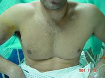
Fig 1: Ecchymoses and asymmetry of the pectoral axillary
fold.
All paients had a
standard delto-pectoral approach in the beach-chair position. A
5-8 cm incision, at the distal end of the delto-pectoral
interval, was utilized for exposure. The avulsed pectoralis
tendon was easily identified. (Fig 2)
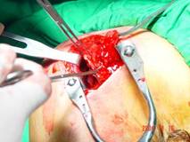
Fig 2:
The avulsed Sternal and Clavicular heads
The insertion site
of the tendon lateral to the bicipital groove was cleaned of any
tendon remnants and roughened to create a bone trough. Three
absorbable anchor sutures (Duet anchor suture Bionx-Linvatec)
® were used to fix the avulsed pectoralis tendon. The absorbable
anchors are inserted into the humeral bone at the intended
fixation site. Two anchor sutures were used to bring the sternal
head deep and proximal to the clavicular head muscle which was
repaired with one suture. (Fig 3)
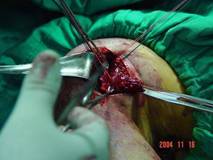
Fig 3: Suture arms through the Sternal and Clavicular heads.
The arm is
slightly adducted and internally rotated, and traction placed on
the sutures arms brings the tendon down to the bone, tying the
suture over the bone trough. (Fig 4)
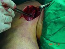
Fig 4: Following repair, reestablishment of the delto-pectoral
groove.
The wound is
irrigated, and closed with subcutaneous absorbable sutures. (Fig
5)
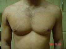
Fig 5:
Symmetry of the pectoral axillary fold restored postoperatively.
Postoperatively, the operated shoulder was immobilized for four
weeks in adduction and internal rotation using
an arm sling.
Patients
were requested to avoid abduction, external rotation, and
resisted internal rotation. They were otherwise allowed passive
motion within these parameters.
Range-of-motion exercises except for abduction and external
rotation were started at four weeks. Six weeks post operatively,
the sling was discontinued,
and abduction and external rotation were initiated. Isometric
exercises were started at two months. Light resistance training
was started at three months and heavy training at four months,
with a return to unrestricted activity at six months.
Results :
At an average
follow-up of fourteen months post repair (twelve to eighteen
months), all patients were satisfied. They had restoration of
the axillary folds with full active range of motion. There was
return to full strength in adduction and internal rotation. All
patients went back to weight lifting and body building sports
activities.
There were no
immediate surgical postoperative complications and no
re-ruptures during the follow up period.
Discussion:
Most, if not all
pectoralis major ruptures occur in males in their second to
fourth decade of life. Most studies have demonstrated
significant improvement in strength following acute repair of
pectoralis major ruptures when compared with non-operatively
treated injuries.2, 3, 11
Nearly two thirds
of these ruptures occur at the tendinous insertion lateral to
the bicipital groove.3
The advocated
technique of tendinous repair is through the use of drill holes
to create bone tunnels at the repair site. Passing sutures
through the bone tunnels with a specific needle that matches the
curve of the drill is somehow cumbersome.12
In our study
we have assessed the use of absorbable anchor sutures to
overcome the above. Duet anchor suture Bionx-Linvatec ® is a
bioabsorbale screw-in suture anchor that is preloaded on a
disposable inserter device with two non-absorbable, braided,
polyester #2 sutures. (Fig 6)
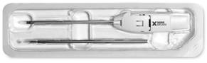
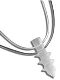
Fig 6: Duet Anchor
Suture
It is manufactured from
Self-Reinforced (96/4D) PLA Copolymer that retains 90% strength
through 20 weeks and completely resorbs over a period of several
years in vivo. The Self-Reinforced Copolymer provides high
initial mechanical strength required for insertion and through
the healing phase (20
weeks) with complete
absorption occurring over several years. The absorption profile
of the Copolymer allows the anchor to gradually loose strength
as the collagen fibers of the repair form and gain strength. The
Copolymer is inert, non-collagenous and non-pyrogenic through
the absorption process.
The insertion is
relatively simple, and provides an excellent pull out strength
which is estimated at 217 N. The material strength eliminates
bioabsorbable eyelet as a failure mode in repair construct, 406
N; 13
this was similar
to our experience with use of these sutures at other sites
(rotator cuff tears, vastus medialis ruptures).
Bal GK and
Basamania CJ have expressed their concern with anchor suture
repair not providing as much broad area of tendon-bone contact
as it was with the routine bone tunnel sutures. This was not the
case in our technique, which is basically due to the presence of
a double suture line that can be utilized over a broader area of
the repaired tendon. We did not have any wound complications
and no tendon repair re-ruptures.
Several studies
have shown a possible correlation between prior steroid use and
subsequent pectoralis major ruptures.2, 14 These
patients should be cautioned concerning healing potential and
overall risk of future medical problems or injuries.
Conclusion:
Pectoralis major
tendon ruptures are uncommon injuries and most physicians have
little experience with the surgical treatment of this injury. We
have presented our experience with use of the absorbable anchor
sutures in the acute repair of tendon ruptures. We feel that
this technique simplifies a cumbersome step of the repair
without altering the postoperative care, rehabilitation, and
complication rate.
Reference :
1. Patissier P.
Maladies des bouchers. Traite des maladies des artisans,
162-165, 1822.
2. Aarimaa V,
Rantanen J, Heikkila J, Helttola I, Orava S. Rupture of the
pectoralis major muscle. Am J Sports Med. 2004; 32:1256-1262.
3. Bak K,
Cameron EA, Henderson IJ: Rupture of the pectoralis major: a
meta-analysis of 112 cases. Knee Surg Sports Traumatol Arthrosc.
2000; 8:113-119.
4. Scott BW,
Wallace WA, Barton MA. Diagnosis and assessment of the
pectoralis major rupture by dynamometry. J Bone Joint Surg Br.
1992; 74:111-113.
5. McEntire JE,
Hess WE, Coleman SS. Rupture of the pectoralis major muscle: a
report of eleven injuries and review of fifty-six. J Bone Joint
Surg Am. 1972; 54:1040-1046.
6. Roi GS,
Respezzi S, Dworzak F. Partial rupture of the pectoralis major
muscle in athletes. Int J Sports Med. 1990; 11:85-87.
7.Hanna CM,
Glenny AB, Stanley SN, Caughey MA. Pectoralis major tears:
comparison of surgical and conservative treatment. Br J Sports
Med. 2001;35:202-206.
8. Liu J, Wu
JJ, Chang CY, Chou YH, Lo WH. Avulsion of the pectoralis major
tendon. Am J Sports Med. 1992; 20:366-368.
9. Quinlan JF,
Molloy M, Hurson B. J: Pectoralis major tendon ruptures, when to
operate. Br J Sports Med. 2002; 36:226-228.
10. Beloosesky
Y, Grinblat J, Weiss A, Rosenberg P, Weisbort M, Hendel D.
Pectoralis major ruptures in the elderly. Clin Orthop. 2003;
413:164-169.
11. Schepsis
AA, Grafe MW, Jones HP, Lemos MJ. Rupture of the pectoralis
major muscle. Outcome after repair of acute and chronic
injuries. Am J Sports Med. 2000; 28:9-15.
12. Bal GK,
Basamania CJ. Pectoralis Major Tendon Ruptures: Diagnosis and
Treatment (Technique). Techniques in Shoulder and Elbow Surgery.
2005; 6(3): 128-134.
13. Barber FA,
Herbert MA, Richards DP. Sutures and suture anchors: update
2003. Arthroscopy. 2003; 19: 985-990.
14. Wolfe SW,
Wickiewicz TL, Cavanaugh JT. Ruptures of the pectoralis major
muscle. An anatomic and clinical analysis. Am J Sports Med.
1992; 20:587-593.
|

