|
Abstract:
Removal of the femoral stem
can be challenging for the orthopaedic surgeon. The decision
between performing an extended trochanteric osteotomy (ETO) or
trying to remove the femoral stem without an osteotomy and
taking the risk of an intraoperative fracture is often hard to
make.
Purpose of the study was
thus to describe our experiences, comparing intraoperative
femoral fractures during stem removal with ETOs in femoral
revision arthroplasties.
45 intraoperative fractures
during revision hip arthroplasty were compared to a collective
of 28 femoral revision arthroplasties. Preoperatively and after
32 months (range, 21.6 - 76 months) the patients were examined
clinically and radiographically. The SF-36 health score, Harris
hip score, a motion- and pain score and the occurrence of
postoperative complications were collected.
Only in ETO patients the
Harris hip score increased significantly (p > 0.01). Results in
pain- and motion scores were better in the osteotomy group.
Outcomes in the SF-36 health score in 3 dimensions were
significantly better in patients with ETOs. No osteosynthesis
related complications occurred in the ETO group, but in 6 (13.3
%) patients in the fracture group (p > 0.01). Re-revision rate
was lower in the osteotomy group. Radiographs showed less bone
resorption, decreased stem subsidence and fewer nonunion in
patients with ETOs.
Well conducted ETOs are
preferable to unplanned femoral fractures during stem removal.
J.Orthopaedics 2007;4(4)e6
index.htm
Introduction:
The number of femoral revision arthroplasties continues to rise
in an ageing population [9]. Removal of the femoral stem can be
a challenge for an orthopaedic surgeon and the decision between
performing a femoral osteotomy or taking the risk of an
intraoperative fracture, being aware that the occurrence of this
complication may lead to disastrous results, is often hard to
make. High fracture rates reported, point out that stem removal
is associated with substantial surgical complications [8, 19].
An intraoperative fracture rate of 7.8 % was reported in
revision Total Hip Arthroplasties (THA) in an review of the Mayo
Clinic Joint Registry [3]. In another study, intraoperative
femoral fractures only occurred in revision hip arthroplasties
at the authors centre [10]. Christensen et al. reported of 10
fractures which occurred exclusively during 159 revision
surgeries (6.3 %) that showed fracture union, but only 6 of 10
had satisfactory postoperative function [6].
Preserving as much bone stock as possible and avoiding any
femoral fracture or crack is crucial in femoral revision
procedure [8]. Extended trochanteric osteotomy (ETO) can
contribute to this, by facilitating cement and stem removal or
implantation of revision components [4, 8, 14, 25, 26].
Additionally, this technique may minimize the risk of
intraoperative fractures and preserves the remaining femoral
bone stock [8, 14, 25, 26]. However, it has also been associated
with new hazards like increased incidence of nonunion, fracture
of the osteotomy fragment and subsidence of the stem [4, 19,
26].
To our knowledge it is not yet clear if a permissive indication
for ETO may lead to better postoperative results than removing
stem and cement from the top of the femur eventually causing
fractures.
Purpose of the present study was thus to describe our
experiences, comparing intraoperative femoral fractures during
stem removal with extended trochanteric osteotomies in femoral
revision arthroplasties in a retroperspective clinical and
radiological study.
Material and Methods :
Between January
1992 and February 2004 593 femoral revision arthroplasties were
performed at our orthopaedic department. All surgical procedures
were carried out by a group of four experienced surgeons. Out of
this number of revision arthroplasties two groups were collected
for the present study. 45 intraoperative fractures were detected
(group I) and compared to a collective of 28 patients which
underwent an ETO during femoral revision arthroplasty (group
II). Preoperatively and after a follow-up period of 32 months
(range, 21.6 months to 76 months) patients were examined
clinically and radiographically.
There were 32 (71.1 %) female and 13 (28.9 %) male patients with
an age range of 38.5 years 88.2 years (mean 67.5 years) in the
fracture group and 10 (35.7%) female and 18 (64.3%) male
patients with an age range of 36.7 years 83.8 years (mean 65.6
years) in the osteotomy group. No statistically significant
differences regarding age at operation were apparent between the
groups. The indication for revision surgery was aseptic
loosening for 37 hips (82.2 %) in the fracture group and for 21
hips (75.0 %) in the osteotomy group, septic loosening for 5
hips (11.1 %) in the fracture group and for 6 hips (21.4 %) in
the osteotomy group and recurrent luxations for 3 hips (6.7%) in
the fracture group and for 1 hip (3.6%) in the osteotomy group.
Patients with ETOs had 1.7 (range, 1 to 4) and in the fracture
group 1.6 (range, 1 to 5) previous surgeries on the involved
hip. No statistically significant differences in indication for
revision surgery and number of previous surgeries were apparent
between the groups.
Classification of fractures:
In group I, fractures during femoral revision arthroplasties
occurred during dislocation, stem or cement removal,
instrumentation or canal preparation. Fractures were graded
using the Vancouver Classification adapted to the intraoperative
scenario [12].
7 (15.5 %) displaced or unstable fractures of the greater
trochanter (A3), 15 (33.3 %) diaphyseal cortical perforations
(B1), 9 (20 %) undisplaced linear cracks (B2) and 7 (15.5 %)
displaced fractures of the midfemur (B3) were monitored.
Furthermore, 5 (11.1 %) displaced fractures of the distal femur
(C3), 1 (2.2 %) cortical perforation of the distal femur (C1)
and 1 (2.2 %) undisplaced linear crack propagating to the distal
femur (C2) occurred. Cerclage wire osteosynthesis was performed
in 34 (75.5 %) hips. For 8 (17.8 %) femoral fractures no
osteosynthesis was needed. In 3 hips (6.7 %) a combination of
plates and cables was brought in. In the osteotomy group,
fragments were reattached with at least 2 cables.
Surgical technique:
We used a standard lateral approach for exposure of the femur
and acetabulum that was extended distally as far as necessary
for the ETO and in case of intraoperative fractures. The ETO
first described by Wagner [22] and then modified by Younger et
al. [25] was performed in all cases in the osteotomy group with
the femoral component still in place. The decision making
process for the ETO involved patient age and condition, implant
design, the use of cement and preoperative radiographs. However,
the final decision was made intraoperatively after the surgeon
assessed bone stock, soft tissue and implant stability.
With an oscillating saw the posterolateral third of the femur
including the complete greater trochanter was cut, beginning
with the anterior lateral osteotomy. Next, the distal end of the
osteotomy was performed with the use of a pencil-tip burr to
minimise stress risers and the risk of fractures. After the
posterior cut, the fragment was retracted anteriorly (Fig. 1).
Only for removal of cemented stems, the distal osteotomy site
was positioned beyond the tip of the stem, in order to
facilitate cement removal. The osteotomy fragment was retracted
always with the attached vastus lateralis and abductors to
retain blood supply and innervation. Bone grafts were used for
osteotomy reinforcement in 7 (25.0 %) cases and for 10 (22.2 %)
fractured femurs. All patients received a dull corundum coated
uncemented titanium-aluminium-niobium alloy long-stem femoral
component (Wagner SL revision stem; Protek AG, Bern,
Switzerland) except for 5 (17.9%) patients in the osteotomy
group and 10 (22.2%) patients in the fracture group who received
a BiCONTACT® Stem (Aesculap, Tuttlingen, Germany) originally
designed for primary THA. In every instance the distal end of
the osteotomy or fracture was bypassed by at least two femoral
diameters [12, 22].
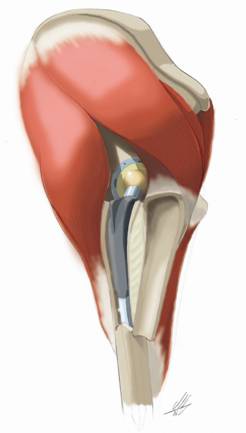
Figure 1 Schematic view on the prosthesis after
extended trochanteric osteotomy. Proximal abductor musculature
and vastus lateralis (not pictured) remain attached to the
fragment.
Figure 2 Improvements of the SF-36 health score in
patients with ETOs and in patients with fractures during femoral
revision arthroplasties.
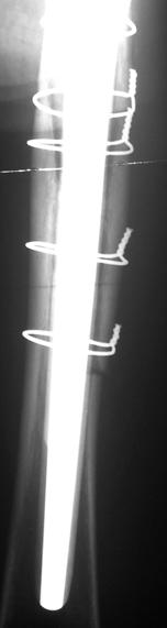
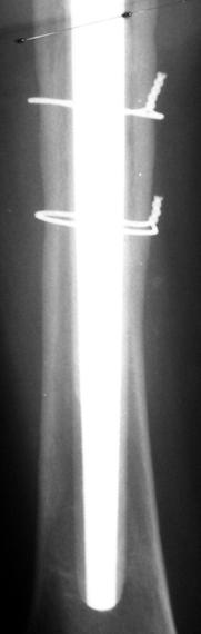
Figure 3a
Three-week postrevision radiograph of a 73-year-old woman shows
distal end of the ETO, a well adapted osteotomy fragment and
good alignment. Figure 3b Follow-up radiograph 6 years
postoperatively shows healed osteotomy without migration and the
ingrown femoral stem in good position
Postoperative treatment:
Depending on the femoral component, acetabular reconstruction,
stability of the hip, and the patients bonestock quality,
postoperative treatment varied. After toe-touch weightbearing
was maintained for 6 weeks, weightbearing was increased to full
bodyweight by 12 weeks. For 6 to 12 weeks, flexion of the hip
was limited to 90°, and active abduction and external rotation
were avoided. Strengthening exercises and passive range of
motion were begun on postoperative day 2.
Preoperatively and after a follow-up period of 32 months (range,
21.6 months to 76 months) patients were examined clinically and
radiographically.
Clinical evaluation:
The patients were routinely followed-up at 3 months, 12 months,
36 months and in intervals of 3 years in our outpatient clinic.
Furthermore patient records were included. For the clinical
follow-up examination the Harris hip score, motion score (visual
analogue scale for flexion of the hip 0 6 points) and pain
score (visual analogue scale 1 10 points) were used. The SF-36
health score (QualityMetric, Incorporated, Lincoln, RI.) was
employed to assess subjective health improvement including 8
multi-item scales: physical functioning, role physical, bodily
pain, general health, vitality, social functioning, role
emotional and mental health, ranging from 0 (maximal symptoms
and poor health) to 100 (no symptoms and excellent health) [23].
In addition, the occurrences of limping and postoperative
complications were assessed.
Radiographic evaluation:
Preoperatively and at follow-up ante-posterior (ap) and axial
radiographs were taken. In 6 patients 3 year follow-up
radiographs were not available. Radiographic evaluation included
the occurrence of fractures or nonunions. Stem migration was
evaluated by measuring the vertical distance between the
shoulder of the prosthesis and the tip of the greater trochanter
on the immediate postoperative ap radiographs and at final
follow-up. Migration of the osteotomy fragment was measured as
the distance between the trochanter fragment and the host bone
on ap radiographs [5]. Since trochanteric fragments may show
migration to craniomedial, axial radiographs were assessed to
quantify the correct distance whenever possible. Implant
alignment was measured by a stem deviation of > 3° from the
femoral longitudinal axis. Osteosynthesis failure, bone
resorption according to Gruen et al. [7] and bone quality
(osteoporosis) were assessed on all radiographs. Bone resorption
affecting less than 50 % of 1 Gruen Zone was rated as not
relevant. The criterion for osteoporotic conditions was, when
the total cortical thickness was less than 25 % of the total
femoral calibre at the midpoint of the shaft [24].
Statistical analysis:
Pooled data of both groups were analysed using 2-sided Students
t-test, paired t-test and Pearsons Chi-Square-Test using SPSS
(version 11.5; Chicago, Illinois). A p value < 0.05 was
considered significant.
Results :
6 patients in the fracture group died 2.6 years after the
operation. No patient in the ETO group was lost to follow-up.
Clinical results:
Results showed an increase in Harris hip scores, pain and motion
scores for all 73 femoral revision arthroplasties. Only patients
with ETOs showed a significant increase in Harris hip scores
(Tab. 1).
Table 1. Clinical scores: Extended trochanteric osteotomies and
femoral fractures in femoral revision arthroplasty
| |
Harris Hip score
[preOP/postOp
|
Pain score
(0-10)
[preOP/postOp]
|
Motion score (0-6)
[preOP/postOp]
|
|
|
|
40/71*
 49/66 49/66
|
|
|
|
In all dimensions of the SF-36 health score, patients with ETOs
had better improvements compared to patients with intraoperative
femoral fractures. Improvements in dimensions Physical
Functioning, Bodily Pain and Vitality were significantly
higher in the osteotomy group (Fig. 2).
Joint luxation occurred in 3 (6.7 %) patients with
intraoperative fractures and once (3.6 %) in the osteotomy group
(not significantly different by Chi-Square-Test). After closed
reduction and change of the femoral head postoperatively, no
more luxations occurred in these patients. No osteosynthesis
related complication was seen in the osteotomy group, but in 6
(13.3 %) patients with intraoperative femoral fractures (p <
0.01): 1 (2.2 %) patient complained about prolonged
postoperative pain over the implanted hardware, in 2 (4.4 %)
cases cable failure occurred during mobilisation, 1 patient
needed refixation of the greater trochanter because of
craniomedial migration and in 2 cases osteosynthesis revision
was needed because of painful hardware. After ETO, 1 (3.6 %)
patient had a transitory lesion of the femoral nerve and 2 (7.1
%) patients had mild Trendelenburg gait, although no
trochanteric nonunion in follow-up radiographs was found. 3 (8.6
%) hips were re-revised in the fracture group (1 nonunion and
subsequent late periprosthetic femoral fracture, 1 aseptic and 1
septic loosening due to nonunion) and 1 (3.6 %) hip needed
re-revision in the osteotomy group (septic loosening and
nonunion).
Radiological results:
Radiographs showed better results in patients with ETOs (Tab.
2), though they were not significantly different by
Chi-Square-Test.
Table 2. Radiographic results: Extended trochanteric
osteotomies and femoral fractures in femoral revision
arthroplasty
| |
Stem
Migration
|
Fragment
Migration
|
Bone
Resorption
|
Osteosynthesis
Failure
|
Nonunion |
| ETO (n28) |
1 (12 mm) |
0 |
1 |
0 |
1 |
| Intraoperative
Fracture (n45) |
3 (15 mm, ± 3 mm) |
|
4 |
2 (cable failure) |
3 |
In all patients except for 1 (3.6 %) in patients with ETOs and 3
(8.6 %) in patients with intraoperative femoral fractures,
osteotomies and fractures achieved union (Fig. 3).
Every implanted stem had excellent alignment (< 3°) in follow-up
radiographs. The number of patients with osteoporotic conditions
was similar with 26 (58 %) patients in the fracture group and 16
(57 %) in ETO patients. In the fracture group, periprosthetic
bone resorption was found in 4 cases: in 1 (2.2 %) case Gruen
Zones 1, 2, 7, 8, 9, and 14 were affected, 1 femur showed bone
resorption in Gruen Zone 1, 1 femur in Zone 5 and in 1 hip Gruen
Zones 2 and 6 were affected. In the osteotomy group, only Gruen
Zone 7 was affected in 1 (3.6 %) patient.
Discussion:
Femoral osteotomies during femoral revision arthroplasty have
always been discussed controversially. Many [8, 14, 25, 26]
support the use of the ETO, although some authors have concerns
with this technique [18, 21] and even give advices to avoid any
femoral osteotomy [15]. However, it is not yet clear if
performing an ETO is preferable to removing the femoral stem
without an additional osteotomy and taking the risk of an
intraoperative fracture.
Purpose of the study was thus to describe our experiences,
comparing intraoperative femoral fractures during stem removal
with ETOs in femoral revision arthroplasties.
In this study, all 73 femoral revision arthroplasties had good
clinical results in Harris hip scores pain- and motion scores,
but solely in patients with femoral osteotomies the increase in
Harris hip score was significant. Results in visual analogue
scale for motion and pain were better in the osteotomy group.
Subjective patient physical and psychological satisfaction with
the operation, represented by the SF-36 health score, showed a
higher increase in all dimensions in patients with ETOs.
However, significantly better increases were only seen in
Physical Functioning and Bodily Pain, as physical
dimensions, and Vitality, as a psychological dimension.
The rate of osteosynthesis related complications was
significantly higher (p < 0.01) in patients with intraoperative
fractures. Furthermore joint luxation was uncommon in patients
with ETOs, whereas it complicated rehabilitation in 3 patients
in the fracture group. In the osteotomy group 2 patients with
Trendelenburg gait were found. Since we did not find a
trochanteric nonunion in these patients, we concur with
Nicholson et al., suggesting a neuromuscular cause [17].
Radiographs showed better results in patients with ETOs.
Periprosthetic bone resorption was found in 10 Gruen Zones in
the fracture group, but only 1 Zone in the ETO group was
affected. Although the overall results in the ETO group were
better compared to the fracture group, we found only few
statistically significant differences between the groups.
Regarding the high number of only minor fractures, the outcome
observed in the fracture group appears to be even poorer
compared to the ETO group. This may demonstrates that small
cracks or perforations during femoral revision might sometimes
be underestimated, especially when previous operations and poor
bonestock make the patient at risk and the bone healing capacity
is compromised. In our opinion undisplaced linear cracks are
easily fixed intraoperatively, but seem to jeopardize prosthesis
ingrowth and stability postoperatively, since these types of
cracks might propagate undetected. However, further studies have
to be conducted to find out why cracks in some patients do
finally become stable and in some they do not.
We note some limitations to our study. Besides the limitations
that come along with a retrospective work, 6 patients in the
fracture group were lost for follow-up after 2.6 years.
We used the SF-36 health score in our study although it is not a
common evaluation tool in orthopaedic surgery, therefore we were
not able to discuss the results of the SF-36 health score with
the literature. However, we believe that we found an
acknowledged and reliable instrument to reflect both physical
and psychological effects [1] in patients after femoral revision
arthroplasties.
Though the two groups are statistically similar, they may have
obtained different treatments intraoperatively. The groups are
possibly different in bonestock damage or in other issues that
led the surgeon to the decision to perform the ETO or to remove
the stem from the top of the femur. Furthermore, it is unknown
if all femurs in the ETO group would have fractured
spontaneously or not. Nevertheless, we believe that the
collectives presented here are well comparable, since the number
of patients with osteoporotic conditions was similar and
indications for revision surgery and age at revision surgery
showed no statistical differences. Even the number of previous
operations, which may be considered as one of the most
determined factors for success in revision surgery, was similar.
This study reflects the problem of intraoperative decision
making in removal of well fixed stems. On the one hand, removing
stem and cement from the top of the femur would be eligible, but
the surgeon has to take the risk of an incalculable fracture. On
the other hand, performing an ETO facilitates stem removal and
may minimize the risk of intraoperative fractures, though it is
an iatrogenic but controllable bone trauma. Addressing this
controversial issue, our results suggest that a more permissive
indication for femoral osteotomy may lead to better
postoperative results, especially in patients with poor bone
stock. Other authors, however, have concerns with the ETO. They
believe that in femoral revision surgery any weakening of the
femur, both intentionally or by accident, compromises femoral
bonestock and may debase postoperative results [21]. In a
recently conducted in vitro cadaver study, the ETO resulted in
a significant reduction of torsional strength and energy
required for fracture [18]. The authors suggest that
rehabilitation should be even more restrictive after revision
total hip arthroplasty with use of the ETO. One author observed
femoral fractures during femoral revision arthroplasty with the
use of the ETO in 12 % (5 of 43 hips) [8]. In a larger series,
25 (20 %) iatrogenic fractures of 122 revision surgeries were
found when this technique was performed [2]. A fracture rate of
10.8 % (18 of 166 hip revisions), propagating from the distal
end of the osteotomy site, were reported in a further study
[14]. Nevertheless, removal of cement, broken implants or well
fixed stems without gaining proper exposure may jeopardize the
femurs integrity, leading to uncontrolled fractures sometimes
even propagating into the supracondylar region [20]. Especially
in patients with poor bone stock, osteoporotic conditions and
previous operations or in septic revision surgery, compromising
the femoral cortex may likely happen. In our clinical
experience, osteosynthesis of an uncontrolled fracture in these
patients poses a great challenge to the surgeon.
The results found in patients with ETOs in our series concur
well with the findings of other authors [2, 8, 14, 25, 26].
Several authors [13, 16] have published techniques to address
the issues of safe cement and stem removal. Wagner [22]
described an osteotomy technique were the greater trochanter and
half of the circumference of the femoral cortex is included.
Popularized by Younger et al. [26], this technique provides
excellent implant, fragment and cement exposure, correction of
proximal femoral deformity, neutral reaming and excellent
acetabular exposure [8, 14, 25, 26]. Besides the excellent
results reported by Paprosky et al. [14, 25, 26] and recently by
Mardones et al. [11] including low complication rates in
fractures, fragment migration, infection, stem subsidence,
instability of the stem and nonunion, we found comparably low
complication rates for stem subsidence and luxation, no
osteosynthesis failure, an acceptable union rate, decreased
postoperative pain and increased range of motion.
Conclusion:
Despite the critical reports found in literature, this study
demonstrates that well conducted extended femoral osteotomies
are preferable to unplanned and uncontrollable femoral fractures
during stem removal.
References :
-
Adachi
JD, Loannidis G, Berger C, Joseph L, Papaioannou A, Pickard L,
Papadimitropoulos EA, Hopman W, Poliquin S, Prior JC, Hanley
DA, Olszynski WP, Anastassiades T, Brown JP, Murray T, Jackson
SA, Tenenhouse A (2001) The influence of osteoporotic
fractures on health-related quality of life in
community-dwelling men and women across Canada. Osteoporos Int
12: 903-908
-
Aribindi R, Paprosky W, Nourbash P, Kronick J, Barba M (1999)
Extended proximal femoral osteotomy. Instr Course Lect 48:
19-26
-
Berry DJ (1999) Epidemiology: hip and knee. Orthop Clin North
Am 30: 183-190
-
Chen WM, McAuley JP, Engh CA, Jr., Hopper RH, Jr., Engh CA
(2000) Extended slide trochanteric osteotomy for revision
total hip arthroplasty. J Bone Joint Surg Am 82: 1215-1219
-
Chin KR, Brick GW (2000) Reattachment of the migrated
ununited greater trochanter after revision hip arthroplasty:
the abductor slide technique. A review of four cases. J Bone
Joint Surg Am 82: 401-408
-
Christensen CM, Seger BM, Schultz RB (1989) Management of
intraoperative femur fractures associated with revision hip
arthroplasty. Clin Orthop Relat Res: 177-180
-
Gruen TA, McNeice GM, Amstutz HC (1979) "Modes of failure" of
cemented stem-type femoral components: a radiographic analysis
of loosening. Clin Orthop Relat Res: 17-27
-
Huffman GR, Ries MD (2003) Combined vertical and horizontal
cable fixation of an extended trochanteric osteotomy site. J
Bone Joint Surg Am 85-A: 273-277
-
Huo MH, Cook SM (2001) What's new in hip arthroplasty. J Bone
Joint Surg Am 83-A: 1598-1610
-
Johansson JE, McBroom R, Barrington TW, Hunter GA (1981)
Fracture of the ipsilateral femur in patients wih total hip
replacement. J Bone Joint Surg Am 63: 1435-1442
-
Mardones R, Gonzalez C, Cabanela ME, Trousdale RT, Berry DJ
(2005) Extended femoral osteotomy for revision of hip
arthroplasty: results and complications. J Arthroplasty 20:
79-83
-
Masri BA, Meek RM, Duncan CP (2004) Periprosthetic fractures
evaluation and treatment. Clin Orthop Relat Res: 80-95
-
Masterson EL, Masri BA, Duncan CP (1998) Surgical approaches
in revision hip replacement. J Am Acad Orthop Surg 6: 84-92
-
Miner TM, Momberger NG, Chong D, Paprosky WL (2001) The
extended trochanteric osteotomy in revision hip arthroplasty:
a critical review of 166 cases at mean 3-year, 9-month
follow-up. J Arthroplasty 16: 188-194
-
Morrey BF, Kavanagh BF (1992) Complications with revision of
the femoral component of total hip arthroplasty. Comparison
between cemented and uncemented techniques. J Arthroplasty 7:
71-79
-
Nercessian OA, Newton PM, Joshi RP, Sheikh B, Eftekhar NS
(1996) Trochanteric osteotomy and wire fixation: a comparison
of 2 techniques. Clin Orthop: 208-216
-
Nicholson P, Mulcahy D, Fenelon G (2001) Trochanteric union
in revision hip arthroplasty. J Arthroplasty 16: 65-69
-
Noble AR, Branham DB, Willis MC, Owen JR, Cramer BW, Wayne
JS, Jiranek WA (2005) Mechanical effects of the extended
trochanteric osteotomy. J Bone Joint Surg Am 87: 521-529
-
Paprosky WG, Martin EL (2002) Removal of well-fixed femoral
and acetabular components. Am J Orthop 31: 476-478
-
Schurman DJ, Maloney WJ (1992) Segmental cement extraction at
revision total hip arthroplasty. Clin Orthop Relat Res:
158-163
-
Turner RH, Emerson RH, Jr. (1982) Femoral Revision Total Hip
Arthroplasty. In: Turner RH, Scheller AD Jr, ed. Revision
Total Hip Arthroplasty. New York, Grune and Stratton: 75-104
-
Wagner H (1989) [A revision prosthesis for the hip joint].
Orthopade 18: 438-453
-
Ware JE, Jr., Gandek B (1998) Overview of the SF-36 Health
Survey and the International Quality of Life Assessment (IQOLA)
Project. J Clin Epidemiol 51: 903-912
-
Watts NB, Harris ST, Genant HK, Wasnich RD, Miller PD,
Jackson RD, Licata AA, Ross P, Woodson GC, III, Yanover MJ, .
(1990) Intermittent cyclical etidronate treatment of
postmenopausal osteoporosis. N Engl J Med 323: 73-79
-
Younger TI, Bradford MS, Magnus RE, Paprosky WG (1995)
Extended proximal femoral osteotomy. A new technique for
femoral revision arthroplasty. J Arthroplasty 10: 329-338
-
Younger TI, Bradford MS, Paprosky WG (1995) Removal of a
well-fixed cementless femoral component with an extended
proximal femoral osteotomy. Contemp Orthop 30: 375-380
|