|
Abstract:
Introduction : Sinding-Larsen Johanssen Syndrome
represents one of the less common causes of anterior knee pain.
It refers to an inflammation of the lower pole of the patella at
the site of origin of the patellar tendon. It is characterized
by tenderness, warmness and swelling over the lower pole of the
patella and also pain with activity. It is considered to be the
junevile equivalent of patellar tendinopathy.
Purpose : To describe a case of an adult male with
Sinding-Larsen Johanssen Syndrome.
Patient and Method : A 34 years old male who came in the
First Aids Department of the Elassona Health Center
complaining of right knee pain with activity, especially when
straightening the leg against force.
Conclusion : Sinding-Larsen Johanssen Syndrome is a very
rare cause of patellofemoral pain, the diagnosis of which is
difficult and it can be confirmed only by the paraclinical
investigations such as X-Rays, Ultrasonography and MRI.
J.Orthopaedics 2007;4(4)e4
Keywords:
Anterior Knee Pain, Patellar Tendinopathy, Patellar
Fragmentation, X-Rays, MRI.
index.htm
Introduction:
The term Sinding-Larsen Johanssen Syndrome refers to an
inflammation of the kneecap ( patella ) at its lowest point.
This is the site of origin of the patellar tendon. There is
traction on the kneecap at this point due to action of the
large, powerful thigh muscle ( quadriceps ). It can be followed
by calcification, ossification or frank inferior pole avulsion
fractures that produce one or more distinct ossicles.
The injury is usually due to repeated stress or vigorous
exercise.
The most common symptoms and signs include tenderness, warmness
and soft tissue swelling over the lower pole of the patella and
also pain with activity, especially when straightening the leg
against force ( such as with stair climbing, jumping, deep knee
bends or weight lifting ) or following an extended period of
vigorous exercise. In more severe cases, appears pain during
less vigorous activity [ 1, 2 ].
Radiographic findings include osseous fragmentation of the
patella and varying amounts and shapes of calcification at the
junction of the patella and the ligament [3 ].
Initial treatment consists of relieving the pain by resting
for a few days and also stretching and strengthening exercises
and modification of activities. Specifically, kneeling, jumping,
squatting, stair climbing and running on the affected knee
should be avoided. Administration of Non-Steroidal
Anti-inflammatory Drugs ( NSAIDs ) may be necessary and in
severe cases a cast is used. In rare cases, operative
debridement of necrotic intratendinous tissue may be needed.
Case Report:
A 34 year
old man came to the First Aids Department of the Elassona
Health Center in Elassona, Larissa, Greece complaining of Right
Knee pain with activity, especially when straightening the leg
against force.
The clinical examination of the patient revealed a
diffuse tenderness and soft tissue swelling over the lower pole
of the affected patella. The rest of the physical examination
has shown no-remarkable findings.
The X-Rays examination of the knee joints revealed
tripartite patella in the right lower limb. Ultrasound
examination have shown fragmentation of the affected patella and
also proximal patellar tendonitis with thickening of the tendon
and heterogenous hypoechogenicity within. Magnetic Resonance
Imaging ( MRI ) Findings involve focal thickening of the right
proximal patellar tendon and also sparing of the anterior
component of the tendon ( typical of chronic patellar
involvement ) and also tripartite patella ( right ).
The diagnosis was: Sinding-Larsen Johanssen Syndrome and the
treatment of the patient was including the following: NSAIDs
were administrated to relieve the pain and also rest and
avoiding vigorous exercise were recommended but there was
no-significant improvement of the patients situation. Thats
why the patient undergone an operative debridement of necrotic
intratendinous tissue and in combination with rest and
application of a small cast, its situation is much better and
after 4-5 months he started an exercise program ( 6 months
duration ) in order to restore strength.
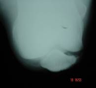 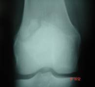
Figure
: A Young male with Sinding-Larsen Johanssen Syndrome
Tripartite Patella
Discussion:
Some of the
most common causes of anterior knee pain are the following:
Chondromalacia Patella, Osteoarthritis of the patellofemoral
joint, Osteochondritis Dessicans, Patellofemoral Dysplasia,
Subluxation of the patella, Dislocation of the patella, Synovial
Plica Syndrome, Tendonitis, Bursitis, Fat Pad Syndrome, Stress
Fractures of the patella, Osgood Schlatter Disease and the
Sinding-Larsen Johanssen Syndrome [ 1, 2, 4, 5, 6, 7, 8 ].
Distinguishing between the possible causes of anterior
knee pain can be difficult as their clinical features can be
similar. Furthermore, some of these conditions may appear
simultaneously as a result of the same biomechanical abnormality
or because of overuse or one may occur first and predispose to
the other.
There are a number of important factors to elicit in the
history of a patient with general presentation of anterior knee
pain. These include the specific location of the pain, the
nature of aggravating activities, the history of the onset of
the pain and any associated clicking or swelling. The type of
activity producing pain also aids diagnosis. The onset of
typical anterior knee pain is often insidious, but it may also
present after an acute traumatic episode.
Disorders such as Chondromalacia Patella, Osteoarthritis,
Osteochondritis Dessicans, Patellofemoral Dysplasia, Subluxation
of the Patella and Dislocation of the Patella are usually
associated with various cartilage damage. Disorders associated
with usually normal cartilage include Tendinitis, Bursitis, Fat
Pad Syndrome, Osgood Schlatter Disease and the Sinding-Larsen
Johanssen Syndrome [ 1, 2, 4, 5, 6, 7, 8 ].
Patellar Tendinitis is an overuse syndrome existing as
tendonitis of the Patellar or Quadriceps tendon at either the
inferior or superior pole of the patella [ 2 ]. Accordimg to the
majority of authors and the international literature, three ( 3
) main sites of tendonitis are described. The first site is at
the attachment of the patellar tendon to the inferior pole of
the patella, second at the insertion of the quadriceps tendon at
the base of the patella and thirdly at the insertion of the
patellar tendon into the tibial tuberosity [ 2 ]. Symptoms
include an insidious onset of low-grade aches aggravated by
deceleration and acceleration forces. Examination exhibits
tenderness and swelling over the tubercle and associated
patellar tendon. Diagnosis is mainly via clinical examination as
the only constant radiographic feature is swelling (greater than
4 mm) over the anterior tibial articular surface. MRI and
Ultrasound can aid in diagnosing the condition. MRI can show
patellar tendon thickening and chrinic tendon tear and
Ultrasound can help distinguish fragmentation [ 3 ].
Sinding-Larsen Johanssen Syndrome is a similar condition which
occurs at the distal pole of the patella. It presents as a
traction apophysitis of the distal pole of the patella secondary
to overuse. Most commonly it is described as the juvenile
equivalent of patellar tendonitis ( but it can also be present
in adults like in our case ) [ 1 ]. The condition presents with
the same symptoms as Osgood Schlatters Disease except at the
opposite location ( Tibial Tuberosity ).
It is
evident from the literature that there are numerous pathological
conditions that can cause patellofemoral pain. The authors
believe that confusion arises due to enormity of conditions that
mimic one another in terms of symptomatology in the anterior
knee region.
History and clinical examination are a crucial factor in
formulating a logical diagnosis, but definite diagnosis can be
based only in the further investigations which can include :
X-Rays, Ultrasound and MRI.
Reference :
1. Greenfield M.A. and Scott W.N. 1994. Patellofemoral Pain. In
Scott W.N.
( Ed ) The Knee. Mosby. St. Louis, Baltimore, Boston, London. pp
: 391-403.
2. David J.M. October 1989. Jumpers knee. The Journal of
Orthopaedic and Sports Physical Therapy. Vol 11 ( 4 ), pp :
137-141.
3. Rask B.P. and Micheli L.J. 1994. The Paediatric Knee. In
Scott W.N. ( Ed ) The Knee. Mosby. St. Louis, Baltimore, Boston,
London. pp : 229-240.
4. Brukner P. and Kahn K. 1994. Clinical Sports Medicine. Mc
Graw-Hill Book Company. pp : 366-400.
5. Merchant A.C. 1994. Extensor Mechanism Injuries :
Classification and Diagnosis. In Scott W.N. ( Ed ) The Knee.
Mosby. St. Louis, Baltimore, Boston, London. pp : 403-409.
6. Calvo R.D, Steadman J.R, Sterling J.C, Holden S.C and Meyers
M.C. 1990. Managing Plica Syndrome of the Knee. The Physician
and Sports Medicine. Vol. 18, (7), pp : 64-74.
7. Johnson D.P., Eastwood D.M and Witherow P.J. 1993.
Symptomatic Synovial Plicae of the Knee. The Journal of Bone and
Joint Surgery. Vol. 75-A, ( 10 ), pp : 1485-1495.
8. Tsirbas A., Paterson R.S. and Keene G.C.R. 1990. Fat Pad
Impingement ; A Missed Cause of Patellofemoral Pain ? The
Australian Journal of Science and Medicine in Sport. Vol. 23, (
1 ), pp : 24-26.
|

