| CASE
REPORT |
|
Spontaneous Rupture Of The Latissimus Dorsi In A Patient Of
Rheumatoid Arthritis |
|
Relwani
J*,Shankaranarayana
S*,Shrivastava R
K*,Brooks C*,Santhakumaran
A**
*
Department
of Trauma & Orthopaedics,
William
Harvey
Hospital
, Ashford,
Kent
**Department
of Radiology,
William
Harvey
Hospital
, Ashford,
Kent
Address for Correspondence:
J Relwani, FRCS (Tr & Orth), FRCS, MS
(Orth), DNB (Orth), MBBS
12 Diamond Estate, 61 Glenburnie Road, London SW17 7DJ.
Email: relwani@hotmail.com |
| |
|
Abstract:
Latissimus dorsi rupture, although uncommon, has been described
in young athletic individuals following trauma. To our
knowledge, a spontaneous rupture of this muscle in a patient of
rheumatoid arthritis has never been reported. Spontaneous tendon
ruptures have been described in rheumatoid arthritis patients on
long term steroid therapy, most commonly the flexor and extensor
tendons of wrist(1), (the achilles tendon, biceps brachii
tendon, and the rotator cuff of shoulder)(2). We describe a case
of spontaneous rupture of latissimus dorsi muscle in a 64 years
old, male rheumatoid arthritis patient on steroid therapy for
duration of 30 years.
J.Orthopaedics 2007;4(4)e25
Keywords:
Spontaneous
latissimus dorsi rupture; rheumatoid arthritis; steroid-induced;
tendon rupture.
Case Report:
A
sixty-four year old man complained of sudden onset of sharp pain
in the left side of his mid-back as he reached out to pick up a
glass of water. He immediately noticed a swelling in the region,
which was followed by bruising. He complained of pain with
movement of left shoulder and on attempting to lie on the
affected side.
He
had no history of trauma or of previous back problems. He was
under treatment for poly articular rheumatoid, and had been on
steroids (prednisolone average dose 6 mg/ day) for 30 years. He had a ruptured right Achilles tendon 6 years
earlier which was treated non-operatively. In addition, he had a
supraspinatus deficient shoulder, but was able to manage
activities of daily living.
Examination revealed extensive bruising over lower third of left side of
back and in the flank (Figure 1). A swelling of 15 x 12
centimetres size was noted with the simultaneous loss of definition of the
posterior axillary fold. The swelling was soft, and became more
prominent when attempting to elicit a contraction of the
latissimus dorsi, with its superior edge moving further distally
by 2.5 cm.
Examination of his shoulder revealed a pre-existing chronic cuff tear
with limitation of active abduction to 70 degrees, external
rotation of 25 degrees and internal rotation to the sacro-iliac
joint.
Functionally patient had difficulty lying down on his back, pushing
himself up from the bed due to pain but was able to manage it
with little discomfort within 72 hours.
He was investigated using an ultrasound scan (Figure 2) and also
underwent magnetic resonance scanning (Figure 3) which confirmed
the presence of the tear in the latissimus dorsi.
The
injury was treated
non-operatively in view of
the limited functional
demands of the patient due
to his co-morbidities, and
the rapid relief of pain
over 72 hours. With the help
of physical therapy for 4
months, he returned to
independent activities of
daily living, but with
persistent weakness of
adduction and internal
rotation of the arm. |
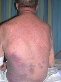 |
| Fig 1: Extensive left flank bruising
and additional skin fold seen in left lower flank. |
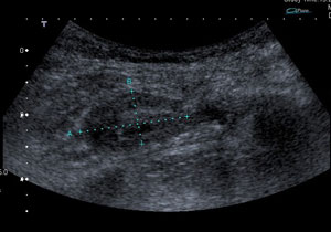 |
| Fig 2: Ultrasound
image confirming a 33mm x 16mm haematoma as a result of the
latissimus dorsi rupture
|
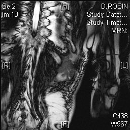 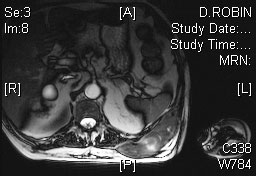 |
| Fig
3: MRI demonstrating high signal changes of haematoma in
left latissimus dorsi muscle in the transverse and coronal
planes |
Discussion:
There is very limited
literature available about latissimus dorsi rupture and the
natural history of the latissimus dorsi rupture is unknown.
Spinner et al(3) described a 38 years old golfer with latissimus
dorsi and teres major rupture , patient had satisfactory results
with conservative management. Kawashima et al(4) described a
case of ruptured latissimus dorsi tendon with a rupture of
pectoralis major post crush injury in a construction worker.
Henry et al(5) described a 42 years old fit skier sustained a
latissimus dorsi rupture which was treated with surgical repair
and re attachment of latissimus dorsi tendon to humerus with
satisfactory results . Butterwick et al(6) describe a 35 years
old, rodeo steer wrestler with a right latissimus dorsi rupture
which was treated conservatively and patient recovered
satisfactorily.
Our patient was a gentleman with underlying rheumatoid arthritis
on prednisolone. This predisposed him to tendon injury, and he
spontaneously ruptured the latissimus dorsi without any obvious
trauma. The diagnosis was confirmed both clinically and on MRI
and US examination. He was treated non-operatively and had
satisfactory function, keeping in mind his original low-demand
status.
This is a rare injury and the first such reported spontaneous
rupture of the latissimus in a rheumatoid patient.
References:
(1)
Ertel AN, Millender LH, Nalebuff E, McKay D, Leslie B. Flexor
tendon ruptures in patients with rheumatoid arthritis. J Hand
Surg [Am ] 1988; 13(6):860-866.
(2) Wanivenhaus
A. [Tendon ruptures in rheumatic patients.]. Z Rheumatol 2007.
(3) Spinner
RJ, Speer KP, Mallon WJ. Avulsion injury to the conjoined
tendons of the latissimus dorsi and teres major muscles. Am J
Sports Med 1998; 26(6):847-849.
(4) Kawashima
M, Sato M, Torisu T, Himeno R, Iwabuchi A. Rupture of the
pectoralis major. Report of 2 cases. Clin Orthop Relat Res
1975;(109):115-119.
(5) Henry
JC, Scerpella TA. Acute traumatic tear of the latissimus dorsi
tendon from its insertion. A case report. Am J Sports Med 2000;
28(4):577-579.
(6) Butterwick
DJ, Mohtadi NG, Meeuwisse WH, Frizzell JB. Rupture of latissimus
dorsi in an athlete. Clin J Sport Med 2003; 13(3):189-191.
|
|
This is a peer reviewed paper Please cite as
:Relwani
J :
Spontaneous Rupture Of The Latissimus Dorsi In A Patient Of
Rheumatoid Arthritis
J.Orthopaedics 2007;4(4)e25
URL:
http://www.jortho.org/2007/4/4/e25 |
|
|

