|
Abstract:
Reconstruction of large bone defect is a major orthopedic
challenge. We are presenting a case of 12 cm long bone defect in
upper tibia following tumor excision. Distal corticotomy and
bone transport using the Ilizarov technique was done to gain
bone regeneration. The residual gap at docking site was bridged
with bone grafting and buttress plating.
Keywords: Ilizarov Technique, bone
defect
J.Orthopaedics 2007;4(4)e19
index.htm
Introduction:
Bone construction to bridge a large bone defect is a challenging
orthopaedic situation. There are several options to bridge a
defect upto 6 cm, but a reliable option for a bone gap of more
than 6 cm is limited1,2 Many times, multiple or
composite operative procedures are needed for a successful
outcome. We are presenting a case report of bone reconstruction
of 12 cm long bone defect of upper tibia which resulted from a
tumor resection. We used the bone transport technique of
Ilizarov1,2,3,4,5 to regenerate bone &
then did buttress plating along with bone graft to achieve a
complete osseous construction.
Case Report :
A 35 year old
lady presented to us on April2004 with a history of swelling in
upper part of tibia of 2 year duration, there was sudden
increase in pain one day before admission. X-ray showed an
expansile osteolytic lesion with trabeculations at upper
metadiaphyseal region. There was also an evidence of
pathological fracture [picture-1]. Radiologically it was
diagnosed as an aggressive aneurysmal bone cyst with
pathological fracture. On 30/04/2004 patient underwent resection
of the tumor and Ilizarov fixation. Distally the resection was
done through normal bone, and proximally marginal excision and
chemical cauterization was done. There was thin slice of
metaphyseal bone proximally and bone gap of 12cm. Histopathology
report was consistent with aneurysmal bone cyst. Bone transport
was started after a latency period of 14 days. Bone transport
was done at a rate of .25mm every 6 hours (1mm per day) [picture-3].
The transport was completed by 4months (August2004), the rings
were retained for another 7 months (March2005) to allow
maturation of the regenerate. The proximal ring became loose
because of pin tract infection.
The Ilizarov
rings were removed at the end of 11 months (March2005). Patient
had non union like situation the upper part. The pin tract
infection subsided after the removal of ring fixator (by
May2005), then the patient was reoperated on 30th May
2005 & bone freshening, buttress plating and bone grafting was
done. There was sound union by November 2005. At 30 month follow
up [picture-4] the patient had 130° knee flexion and
complete extension. There was terminal restriction of ankle
movements. There was no functional disability. No recurrence of
the tumor.
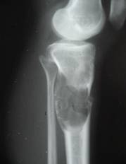 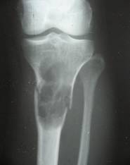
Fig.1
Fig.2
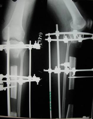
Fig.3
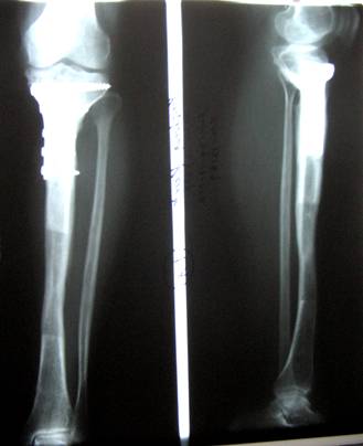
Fig.4
Discussion:
The osseous reconstruction of a large segmental defect in a long
bone requires an surgical endeavour which is technically
difficult, time consuming and often leads to multiple operative
procedures1.The whole procedure is physically,
psychologically and economically demanding to the patient &
it has no guarantee of a satisfactory outcome. For a segmental
defect of more than 6 cm of tibia, what is preferred is
vascularised fibular graft and ring external fixator using
Ilizarov technique1,2. The first option necessitates
a vascular sacrifice, may lead to a graft fracture & requires
enough bone at both side for fixation. This procedure was
abandoned in our case once it was found peroperatively that
proximal metaphysical part was too small to hold the fibular
graft.
Bone transport with the Ilizarov technique is a favoured method
of treating a segmental defect of tibia2 We have done
distal corticotomy with distraction osteogenesis using the
Ilizarov technique1,2,3,4.5 to regenerate a bone
segment of 12 cm length. Compared to other methods, it is
usually safer, less expensive and simpler to perform2.
However, like other methods, it has its list of
complications and drawbacks. It is inconvenient to the patient
and requires patients compliance and cooperation. It has a long
fixator time. In our case it was 11 months. There was pin tract
infection at 2 different occasions which required antibiotics &
dressings. The concave under-surface of the proximal
metaphysical part was filled up with fibrous tissue by the time
, the transport disc reached there. The leading edge of the
transport disc was also covered with fibrocartilagenous cap.
This necessitated the excision of fibrous tissue, bone grafting
and buttress plating. The whole tibia was fully reconstructed
clinically and radiologically at 17 months after initiation of
treatment.
Ilizarov technique is a safe & reliable procedure to bridge a
large bone defect like this case.
Reference :
1. Keating JF, Simpson AHRW, Robinson CM The management of
fractures with bone loss Journal of Bone & Joint Surgery [Br]
2005;87-B:142-150
2. Sen C, Eralp L, Gunes T, Erdem M, Ozden VE, Kocaoglu MAn
alternative method for the treatment of nonunion of the tibia
with bone loss Journal of Bone & Joint Surgery [Br]
2006;88-B;783-789
3. Goulet JA & Hak DJ Nonunion & malunions of the tibia
In:Chapman Michael W,Chapmans Orthopaedic Surgery,Vol 1 Third
Edition Lippincott Williams & Wilkins, 2001 977-999
4. Green SA Management of fractures, nonunions, and malunions
with Ilizarov techniques In:Chapman Michael W, Chapmans
Orthopaedic Surgery,Vol 1 Third Edition Lippincott Williams &
Wilkins ,2001 1001-1107
5.Ilizarov GA Lengthening of upper & lower limb segments In:
Ilizarov GA Transosseous Osteosynthesis Spring-Verlag 287-328
|

