|
Abstract:
Purpose
Lumbar spinal stenosis is the narrowing of the osteoligamentous
vertebral canal causing compression of the neural elements
within the spinal canal, the lateral recesses, or intervertebral
foramina. Surgery aims to decompress the nervous structures,
particularly the nerve roots, without compromising vertebral
stability. Different surgical modalities have been implicated
with different success rates. The purpose of this study is to
assess the clinical and functional outcome after multiple
laminotomy in treatment of lumbar spinal stenosis.
Methods
Fifty-six patients with lumbar canal stenosis were operated on
using the multiple laminotomy technique after adequate
unsuccessful conservative treatment. Far lateral superior and
inferior laminotomies limited to one half of the superior lamina
and one quarter of the inferior lamina together with the
intervening ligamentum flavum were performed. Attention was
given to lateral recess and root canal decompression. In 8
patients multiple laminotomies failed to achieve adequate
decompression due to absolute stenosis, and were excluded from
the results. Patients were followed up for a mean period of
27.63 ± 10.56 months and assessed according to the Japanese
Orthopaedic association (JOA) score.
Results
Satisfactory (excellent and good) results were obtained in 87.5%
of the patients with mean recovery rate of JOA score of 65% (P <
0.000). Six minor dural tears without residual neurological
signs were recorded. Permanent neurological (root) injury was
recorded in only 2 patients.
Conclusion
Results of the present study proved multiple laminotomy
technique to be the procedure of choice for mild to moderate
developmental and degenerative stenosis.
keywords:
Lumbar spinal stenosis; Interlaminar
decompression; Multiple laminotomy
J.Orthopaedics 2007;4(4)e14
index.htm
Introduction:
Lumbar spinal stenosis (LSS) is the narrowing of the
osteoligamentous vertebral canal causing compression of the
neural elements within the spinal canal, the lateral recesses,
or intervertebral foramina. 1,2,3 The narrowing may
be limited to a single motion segment or more diffuse, spanning
two or more segments.4 LSS can be classified in
several ways based on the anatomic location of the narrowing or
on the aetiology.5,6 Degenerative spinal stenosis is
the most common subtype found in patients seeking medical care.6
Developmental stenosis, on the other hand, presents earlier in
age with similar clinical findings but with multilevel
involvement and fewer degenerative changes.7 The
clinical hallmark finding of lumbar stenosis is neurogenic
claudication presenting as intermittent pain or parasthesia in
the legs brought on by spinal extension, and classically
relieved by flexion.4,7 Several studies have
confirmed the effectiveness of both non surgical treatment and
surgical decompression for the management of LSS but there is
also a general agreement that the more severe the stenotic
symptoms and signs, the greater the role of surgery.2,4,8-10
Traditionally surgical treatment of spinal stenosis is carried
out through a decompressive laminectomy with a limited
facetectomy. Some authors recommend a less invasive approach
using unilateral laminotomy, bilateral laminotomy
(fenestration), bilateral partial laminectomy, a unilateral
approach for bilateral decompression, and interspinous process
decompression.2,9,11-14 These limited approaches were
designed to decrease patient morbidity with faster
rehabilitation; limit surgery to the pathologic area only;
decrease postoperative spinal instability; and potentially avoid
the need for fusion.13 The aim of the present study
was to assess the clinical and functional outcome of multiple
laminotomy technique in the treatment of LSS.
Material and Methods :
Fifty-six patients with symptomatic LSS were the
subject of this prospective study between January 2003 and June
2005. There were 34 males and 22 females ranging in age from 21
to 68 years with a mean of 58.61 ± 10.56 years. All patients had
a previous unsuccessful adequate conservative treatment.
Patients with obvious spinal instability or any previous spinal
surgery were excluded from this study. Detailed history taking,
with standardized general and neurological assessment of the
patients were performed. Patients were clinically evaluated
using the score rating system of the Japanese Orthopaedic
Association (JOA Score) shown in table 110. Patients
were radiologically investigated using plain X-ray, CT scan, and
MRI. The central canal was considered relatively stenotic when
the mid-sagittal diameter was between 10 12 mm or when the
cross sectional area of the dural sac was 80 130 mm2.
Absolute stenosis was considered when the mid-sagittal diameter
was less than 10 mm or the cross sectional area of the dural sac
less than 80 mm2. The root canal was considered
stenotic when its diameter was less than 3 mm.19
All patients were subjected to surgical decompression using
multiple laminotomy technique without spinal fusion through the
standard posterior approach with the patient in the flexed prone
position. The bone from the inferior aspect of the cranial
lamina and, to a minimal degree, from the superior aspect of the
subjacent lamina was resected, and subsequent flavectomy was
performed to expose the spinal canal. The medial aspect of the
facet joint was resected to decompress the lateral recess. The
spinous process, the supra- and interspinous ligaments, and a
substantial portion of the lamina were preserved.9,13,15
|
Table 1: JOA scoring system for
low back pain10
|
|
I. Subjective symptoms |
|
(9 points) |
|
A. Low back pain |
|
|
|
a. None |
3 |
|
|
b. Occasional mild pain |
2 |
|
|
c. Frequent mild or
occasional severe pain |
1 |
|
|
d. Frequent or continuous
severe pain |
0 |
|
|
B. Leg pain and / or
Tingling |
|
|
|
a. None |
3 |
|
|
b. Occasional slight
symptoms |
2 |
|
|
c.
Frequent slight or occasional severe symptom |
1 |
|
|
d. Frequent or continuous
severe symptom |
0 |
|
|
C. Gait |
|
|
|
a. Normal |
3 |
|
|
b. Able to walk > 500 meters
although in pain, tingling, and / or muscle weakness |
2 |
|
|
c. Unable to walk > 500 meters
owing to pain, tingling, and / or muscle weakness |
1 |
|
|
d. Unable to walk > 100 meters
owing to pain, tingling, and / or muscle weakness |
0 |
|
|
II. Clinical signs |
|
(6 points) |
|
A. Straight-leg-raising test
(including tight hamstrings) |
|
|
|
a. Normal |
2 |
|
|
b. 30 70 degrees |
1 |
|
|
c. < 30 degrees |
0 |
|
|
B. sensory disturbance |
|
|
|
a. None |
2 |
|
|
b. Slight disturbance
(not subjective) |
1 |
|
|
c. Marked disturbance |
0 |
|
|
C. Motor disturbance |
|
|
|
a. Normal (Grade 5)
|
2 |
|
|
b. Slight weakness (Grade
4) |
1 |
|
|
c. Marked weakness (Grade
3 0) |
0 |
|
|
|
|
|
|
|
|
|
|
III.
Restriction of ADL (Activities of Daily Living) |
|
ADL |
Severe restriction |
Moderate restriction |
No restriction |
|
|
a. Turn over while lying |
0 |
1 |
2 |
|
|
b. Standing |
0 |
1 |
2 |
|
|
c. Washing |
0 |
1 |
2 |
|
|
d.Leaning forwards |
0 |
1 |
2 |
|
|
e. Sitting (about 1 hour) |
0 |
1 |
2 |
|
|
f. Lifting or holding
heavy objects |
0 |
1 |
2 |
|
|
g. Walking |
0 |
1 |
2 |
|
|
IV. Urinary bladder function |
|
|
|
(-6 points) |
|
a.Normal
0 |
|
|
|
|
|
b.Mild dysuria -3 |
|
|
|
|
|
c.Severe dysuria
-6 |
|
|
|
|
|
* Incontinence |
|
|
|
|
|
*Urinary retention |
|
|
|
|
Intraoperatively, multiple laminotomy procedure failed to
adequately decompress the neural elements due to tight
(absolute) canal stenosis in 8 out of the 56 patients included
in the present study. Hence, in the same operative sittings, the
decompression was extended to total laminectomy, and these
patients were excluded from the study.
Forty-eight patients were followed up for a mean period of 27.63
± 10.56 (11-45), and the final clinical and functional results
were calculated by the formula (Postoperative JOA Score
Preoperative JOA Score / (Total Score Preoperative JOA Score)
x 10010. The result was rated as excellent when JOA
score ranged between 100% - 81%, good when score ranged between
80% - 66%, Fair when score ranged between 65% - 50% and poor
when the score was below 50%. Excellent and good results were
considered a satisfactory result while fair and poor results
were considered unsatisfactory. Data were statistically analyzed
by the SPSS data processing program for windows using the
Students t-test.
Results :
Forty-eight patients with LSS were subjected to spinal
decompression using multiple laminotomy technique. There were 30
males and 18 females (1.7:1) with a mean age of 61.96 ± 5.29 (47
68) years. They were followed up for a mean period of 27.63 ±
10.56 (11-45) months. The clinical symptoms and signs of LSS
among patients are summarized in table 2. All patients underwent
CT and/or MRI imaging evaluation for the spinal stenosis.
Twenty-four patients (50%) had spinal stenosis at the L3L4
level, 18 patients (37.5%) at L4L5 level, and 6 patient (12.5%)
at L2L3 level. Forty patients had one level and 6 patients had
two levels of segmental involvement. Three-segmental involvement
was noted in 2 patients. Evidence of disc herniation was present
in 40 patients mostly at L3-4 and L4-5 levels. Pathologically,
there were 16 patients with developmental stenosis, 18 with
degenerative stenosis, and 14 with combined stenosis.
Intraoperative findings are summarized in table 3. Fourteen
patients (29.2%) underwent bilateral laminotomy alone. Ten
patients (20.8%) had discectomy in addition to laminotomy, 8
patients (16.7%) had a combination of laminotomy and facetectomy,
and 16 patients (33.3%) had a combination of laminotomy,
discectomy and facetectomy. Twelve minor operative complications
were recorded (25%) and included superficial wound infection in
2 patients, urinary tract infection in 2 patients, and dural
tears without residual neurological signs in 8 patients. Major
complications were noted in only 2 patients with permanent
neurological (root) injury. No deep wound infections or thrombo-embolic
complications occurred in the present study.
|
Table 2: The clinical symptoms
and signs of LSS in the studied patients
|
|
Clinical symptoms and signs Number of patients |
No
of patients |
% |
|
Low
back pain |
48 |
(100%) |
|
Intermittent claudication |
48 |
(100%) |
|
Neurological impairment |
48 |
(100%) |
|
Motor
impairment alone |
0 |
(0%) |
|
Sensory impairment alone |
14 |
(29%) |
|
Reduction in reflexes (general) |
8 |
(17%) |
|
Decrease ankle reflex alone |
8 |
(17%) |
|
Decrease patellar reflex |
2 |
(4%) |
|
Urine
incontinency |
2 |
(4%) |
|
Anal
incontinency |
0 |
(0%) |
|
Intact SLR examination |
0 |
(0%) |
|
Positive SLR test (unilateral) |
12 |
(25%) |
|
Positive SLR test (bilateral |
36 |
(75%) |
|
Table 3: Intraoperative
findings
|
|
Operative findings |
No of patients |
% |
|
Obliteration of the
inter-laminar space |
38 |
79.2 |
|
Thickening of lamina |
42 |
87.5 |
|
Hypertrophied medially
displaced facet |
18 |
37.5 |
|
Hypertrophy of the ligamentum
flavum |
48 |
100 |
|
Sparsity of epidural fat |
48 |
100 |
|
Stenosis
Central stenosis
Recess stenosis
Root canal stenosis |
48
6
18 |
100
12.5
37.5 |
|
Soft disc herniation |
40 |
83.33 |
|
Reappearance of epidural fat |
48 |
100 |
|
Return of dural pulsation |
48 |
100 |
The mean JOA score was 7.5 ± 0.8 preoperatively and 12.3 ± 0.9
at the last follow up, with mean recovery rate of 65% (P <
0.000). The overall obtained satisfactory results were 87.5%
(37.5% 18 patients excellent, and 50% 24patients good),
while there were 12.5% unsatisfactory results (8.33% 4
patients fair, and 4.17% 2 patients poor).
Clinical
cases
Case № (1)
A male patient aged 47 years presented with low back pain;
referred bilateral legs pain and intermittent neurogenic
claudication limiting his working activity for one-year
duration. Pain increased by walking and standing in extension
and relieved by sitting. Neurological studies revealed positive
straight leg raising test on the right side at 60o,
there were hyposthesia and motor weakness of L5 dermatome. The
claudication distance was 300 meters. The pre-operative JOA
Score was 19 points.
Radiological studies revealed moderate central canal
stenosis at L4 level (mid-sagittal diameter of 11.5 mm), right
postero-lateral L4 L5 disc prolapse compressing the right L5
nerve root and thickened ligamentum flavum (Fig. 1 A, B, C & D).
The operative procedure for decompression was L4 laminotomy and
L4 L5 discectomy. Intra-operative findings revealed thick
sclerosed laminae, narrow interlaminer space, stenosis of
central spinal canal at L4 level, prolapse of L4-L5
intervertebral disc, sparsity of epidural fat and absent dural
pulsation. There were no intra-operative or post-operative
complications.
The duration of follow up was 25 months. Neurogenic claudication
and leg pain was completely relived. The final JOA score was 28
point. The overall improvement rate was 90% (excellent outcome).
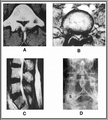
Figure 1: Preoperative (A, B & C) and postoperative (D)
radiographs of case No 1.
Case № (2)
A housewife aged 50 years, presented with low back pain,
referred right leg pain and neurognic intermittent claudication
of 2 years duration. Leg pain increased by walking and prolonged
standing with back extension and relieved by sitting.
Claudication distance was 500 meters. Neurological examination
was unremarkable. The pre-operative JOA score was 17 points.
Radiological studies revealed multilevel moderate central canal
stenosis at L3, L4 and L5 level (mid-sagittal diameter 11.5 mm),
thickened ligamentum flavum and disc bulge at L4-5 and L5-S1
level. (Fig. 2 A, B, C, D & E)
The operative procedure for decompression was multiple
laminotomy at L3, L4 and L5 levels. The intra-operative findings
were thick sclerosed laminae, narrow interlaminar space,
sparsity of epidural fat, thickened ligamentum flavum, absent
dural pulsation and stenosis of central spinal canal. There were
multilevel disc bulge at L3-4, L4-5 and L5-S1. Post-operative
superficial wound infection occurred and resolved by systemic
antibiotics.
The duration of follow up was 24 months. The final assessment
revealed marked improvement of symptom and work ability. The
final JOA score was 26 points. The overall improvement rate was
75% (good outcome).
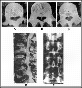
Figure 2: Preoperative (A, B, C & D) and postoperative
(E) radiographs of case No 2.
Discussion:
Forty-eight patients suffering from stenosis of the lumbar
spinal canal were the subjects of this study. All patients were
subjected to previous unsuccessful conservative treatment.
Patients with obvious spinal instability or any previous spinal
surgery were excluded from this study. At the end of follow up
period, the surgical outcome was evaluated both clinically by
JOA scoring system and radiologically.
Patients of this study were 30 males and 18 females, with male
to female ratio 1.7:1 respectively with a mean age of 48.2 ±
11.094 years. This is in consistence with the findings of Fahy
and Nixon1, Fritz et al4, and Benoist8.
The paucity of females suffering from lumbar canal stenosis can
be attributed to less abuse of their spines; their canals may be
generally wider, variation in the vascular anatomy of the female
pelvis, or hormonal factors8. Almost all authors
agree that lumbar canal stenosis occurs in middle and old age
when degenerative changes supervene, reaching their maximum
prevalence in the 5th and 6th decades of life and account for
the late onset of symptoms in most patients; they believed that
the so-called developmental stenosis remains asymptomatic until
the critical reserve space for the enclosed neural elements
becomes compromised by structural changes associated with aging
and trauma1,3-5,8.
All patients included in this study were suffering from low back
pain and unilateral or bilateral neurogenic claudication at the
onset of presentation. These results are consistent with those
of Fahy and Nixon1, Spivak3, Fritz et al.4,
Benoist8, Postacchini et al.9, and
Eule et al.11, who reported an incidence of 85 100
% of different types of complaints. On the contrary, Thome et
al.13, reported an incidence of 6-19% of complaints.
Neurogenic claudication was
present in all of our patients. Mixed sensory deficit (numbness
and hyposthesia) and motor weakness were the commonest
claudication symptom experienced by our patients. Motor weakness
alone was not experienced by any of our patients. These results
are not consistent with the findings of Thome et al13
where claudication leg pain occurred in 93% of their patients
while sensory deficit occurred in 63% and motor weakness in 43%.
Results of the present study are also in contrast with those of
Postacchini et al9 where motor weakness was the
commonest claudication symptom (80.5%) followed by leg pain
(47.4%), and sensory deficit was the least frequent (45.3%).
Intraoperatively, the narrowing of the lumbar canal was found to
be due to combinations of ligamentum flavum hypertrophy (100%),
thickening of the lamina (86.5%), and facetal hypertrophy
(37.5%). Concomitant disc prolapse contributed to narrowing of
the lumbar canal in 83.33% of patients. These findings are in
accordance with findings of different studies1,2,8,10,11,13
who found that degenerative changes in the facet joints and
intervertebral discs, as well encroachment upon the canal by
hypertrophied ligamenta flava were the commonest abnormalities
encountered with in their patients.
All patients had central stenosis, 37.5% had associated root
canal stenosis and 12.5 % had associated lateral recess stenosis.
This coincides with findings of Benoist8, Thome et
al.,13 and Yamazaki et al.,15 who found
that central canal stenosis is rarely an isolated occurrence,
and usually, a variable degree of lateral canal narrowing
coexists with central stenosis and that the nerve root canals
should be the area of primary surgical interest.
During operation, the highest incidence of stenosis was found to
be at L4 and L3 levels (about 50% of cases), followed by L4-5
level (18 pattients), and L2-3 level in 6 patients. These
findings coincide with those of different studies2,9,11,13,15
Decompression at a single level was done for 83.33% of
cases, while two level decompressions were done for 12.5% of
cases. Three level decompressions were done for 4.17% of case.
These findings are not consistent with those of Shabat et al.5,
Postacchini9, Taniguchi et la.10, Eule11,
and Thome et al.13, who performed two or three level
decompressions in 37 45 % of their patients.
Complications in the present study were represented
intraoperatively by dural tears (16.7%) and postoperatively by
superficial wound infection in 4.17% of cases and urinary tract
infection in 4.17% of cases. In other studies the dural tears
range between 0.3% -13%2,11,13. However, these tears
didnt affect the postoperative result. Benoist8
declared that one of the surgical problems in lumbar canal
stenosis is adherence of the dura along the deep medial portions
of the facets at the areas of greatest compression.
The postoperative assessment of relief using JOA score revealed
overall 87.5% satisfactory results (37.5% excellent and 50%
good). The overall unsatisfactory results were 12.5% (8.33% fair
and 4.17% poor). These results are in accordance with those of
Haba et al.2, Eule et al.11, and Thome et
al.13, who achieved satisfactory results in 78-84% of
their patients. The present study showed a significant
improvement in the unrestricted walking distance at the latest
follow up examination compared with per-operative distance (claudication
distance) in all patients. This finding coincides with that of
Eule et al.13.
Satisfactory results were obtained in younger age group and in
patients with short duration of back pain and claudication
symptoms when compared with unsatisfactory results. These
findings are consistent with those of Fritz et al.4
and Eule et al.11, who found that long-standing
compression on the nerve roots would result in irreversible
damage to the nervous tissue and worsen the expected
post-operative outcome. In accordance with Fritz et al.4,
the association of disc prolapse with stenosis had a higher
proportion of excellent results than those with stenosis alone,
and the group of patients with preoperative neurological
deficits had better results than the groups without deficits.
Conclusion:
Of the 48 patients included in this study, satisfactory results
were obtained in 87.5% of cases. The best results were in the
younger age group. Claudication symptoms predicted more
satisfactory results. The longer the claudication distance, and
the shorter the duration of symptoms, the better was the
prognosis. Disc prolapse and discectomy was consistent with more
satisfactory results. Claudication pain, and claudication
weakness were the commonest symptoms to be improved
postoperatively, while low back pain was the least. These
results indicate that multiple laminotomy is an effective method
and the treatment of choice for developmental, degenerative, or
combined lumbar canal stenosis with preservation of vertebral
stability.
Reference :
- Fahy D, Nixon JE. Lumbar Spinal Stenosis. Current
Orthopaedics 2001; 15: 91-100.
- Haba K, Ikeda M, Soma M, Yamashima T. Bilateral
Decompression of Multilevel Lumbar Spinal Stenosis Through a
Unilateral Approach. Journal of Clinical Neuroscience 2005;
12: 169-71.
- Spivak JM. Current Concepts Review. Degenerative Lumbar
Spinal Stenosis. The Journal of Bone and Joint Surgery (Am)
1998; 88-A: 1053-66.
- Fritz JM, Delitto A, Welch WC, Erhard RE. Lumbar Spinal
Stenosis: A review of Current Concepts in Evaluation,
Management, and Outcome Measurements. Arch Phys Med Rehabil
1998; 79: 700-8.
- Shabat S, Leitner Y, Nyska M, Berner Y, Fredman B,
Gepstein R. Surgical Treatment of Lumbar Spinal Stenosis in
Patients Aged 65 Years and Older. Archives of Gerontology and
Geriatrics 2002; 35: 143-52.
- Vo AN, Kamen LB, Shih VC, Bitar AA, Stitik TP, Kaplan RJ.
Rheabilitation of Orthopedic and Rheumatologic Disorders. 5.
Lumbar Spinal Stenosis. Arch Phys Med Rehabil 2005; 86: 69-76.
- Singh K, Samartzis D, Vaccaro AR, Nassr A, Andersson GB,
Yoon ST, et al. Congenital Lumbar Stenosis: A Prospective
Control-Matched, Cohort Radiographic Analysis. The Spine
Journal 2005; 5: 615-22.
- Benoist M. The Natural History of Lumbar Degenerative
Spinal Stenosis. Joint Bone Spine 2002; 69: 450-7.
- Postacchini F, Cintti G. The Surgical Treatment of Central
Lumbar Stenosis: Multiple Laminotomy Compared with Total
Laminectomy. The Journal of Bone and Joint Surgery (Br) 75-B
1993; 385-92.
- Taniguchi S, Yamamoto H. Decompression Surgery for Lumbar
Spinal Stenosis. Current Orthopaedics 1999; 13: 184-90.
- Eule JM, Breeze R, Kindt GW. Bilateral Partial Laminectomy:
A Treatment for Lumbar Spinal Stenosis and Midline Disc
Herniation. Spine 1999; 52: 329-38.
- Rao RD, Wang M, Singhal P, McGrady LM, Rao S. Intradiscal
Pressure and Kinematic Behavior of Lumbar Spine After
Bilateral Laminotomy and Laminectomy. The Spine Journal 2002;
2: 320-6.
- Thome C, Zevgaridis D, Leheta O, Bazner H,
Pockler-Schoniger C, Wohler J, et al. Outcome After
Less-Invasive Decompression of Lumbar Spinal Stenosis: A
Randomized Comparison of Unilateral Laminotomy, Bilateral
Laminotomy, and Laminectomy. J Neurosurg: Spine 2005; 3:
129-41.
- Zucherman JF, Hsu JY, Hartjen CA, Mehalic TF, Implicito
DA, Martin MJ, et al. A Multicenter, Prospective, Randomized
Trial Evaluating the X STOP Interspinous Process Decompression
System for the Treatment of Neurogenic Intermittent
Claudication. Spine 2005; 30: 1351-8.
- Yamazaki K, Yoshida S, Ito T, et al. Postoperative Outcome
of Lumbar Spinal Canal Stenosis After Fenestration:
Correlation With Changes in Intradural and Extrdural Tube on
Magnetic Resonance Imaging. Journal of Orthopaedic Surgery
2002; 10: 136-43.
|

