|
J.Orthopaedics 2007;4(3)e9
Keywords:
Hip; Arthroscopy
Introduction:
Arthroscopy
of the hip is now an established means for diagnosing and
treating a variety of intra-articular pathology. It offers the
benefits of being a minimal invasive procedure with short
rehabilitation, minimal complications and allows for
opportunities for future surgical interventions.1
Better understanding of the arthroscopic anatomy, operative
techniques and potential complications combined with proper
patient selection have widened the scope for hip arthroscopy.
Our
study was undertaken to look at indications, findings,
complications and management outcomes from hip arthroscopy
performed at our centre over a two year period. The aim of this
paper is also to state the common indications, surgical methods,
complications and post operative rehabilitation following hip
scope.
Material and Methods :
A
retrospective case series study was conducted at The Royal
Orthopaedic Hospital,
Birmingham
involving all patients who underwent hip arthroscopy under the
senior author (M.A.G). All the procedures were performed from
April 2004 to Nov 2006 and were followed up for a minimum period
of six weeks. A total of 35 hips were scoped in thirty five
patients (15 male and 20 female). All the patients were assessed
on basis of history of symptoms, physical examination and
radiological investigations in the form of a CT scan or MRI
scan. The average age was 32 years (range11-55 years). 18 right
and 17 left hips were evaluated.
The
procedure was done as a day case surgery under general
anesthesia in all cases. Patients were positioned supine and a
standard fracture table was used with the hip in
mild
abduction and neutral rotation. A well padded lateral perineal
post functioned as a fulcrum to aid in joint distraction. A 16G
spinal needle, passed anteriorly, was utilised to break the
vacuum under the image intensifier. Joint distraction of 5 to
10mm was obtained through traction on the extremity and
confirmed on fluoroscopy. Standard antero-lateral and
postero-lateral portals were then dilated over a guide wire and
either a 300
or 700
arthroscope was introduced into the joint. Hip distraction
was aided by distension with saline using a pump infusion
system. Average duration of the procedure lasted 30-45 minutes
and 20 ml of 0.5% Marcain was instilled into the joint and
infiltrated along the portal sites. Patients were taken off the
traction table immediately after and the perineum looked for any
pressure sores.
Post
operative rehabilitation allowed for full weight bearing with
crutches as tolerated with advice about gentle range of movement
exercises. Patients were discharged home the same day and an
initial follow up appointment made for in six weeks.
Complications and treatment outcomes were assessed during these
and subsequent follow up visits.
Results :
The
average age of our study population was 32 years, with the
youngest being an 11 year old boy presenting with a post
traumatic osteochondral loose body. The oldest subject was a 55
year lady with a degenerate hip. (Figure 1)
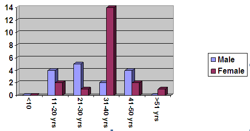
Figure 1. Age and sex distribution of the study
group
Apart
from pain, clicking and snapping were the predominant symptoms.
All the patients were clinically assessed, operated and followed
up by the senior author (M.A.G), but, except for one patient,
they all had been treated previously by one or more orthopedic
surgeon. Preoperative imaging in the form of a CT scan (plain or
contrast enhanced) or MRI scan was obtained in all patients.
A
working diagnosis was established based on the clinical
presentation and imaging findings. In seven patients neither a
clinical nor a radiological diagnosis could be made and were
diagnosed as idiopathic painful hip. Hip arthroscopy was carried
out for removal of loose bodies in six patients, of which one
was diagnosed as intra-articular osteochondroma. Of the rest,
four were post traumatic and one in a perthetic hip. There were
fifteen patients with a positive radiological signs which
included labral fraying or tear (six patients), sub-chondral
cysts or signal changes (six patients), two patients with
synovitis and one patient with osteochondritis dessicans. Three
patients underwent arthroscopic washout for an osteoarthritic
hip. Diagnostic arthroscopy to asses the articular surfaces for
planned pelvic and femoral osteotomies was the indication in
four patients. (Figure 2)
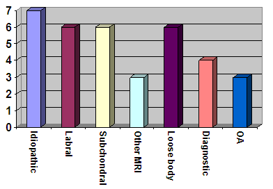
Figure 2. Indications for Hip Arthroscopy
A standard procedure for performing hip scope, as described
earlier, was adopted in all cases. Access to the joint was
possible without fail in all hips. The duration of the procedure
was from 30 to 45 minutes. In ten hips normal looking articular
surfaces and labrum was observed. Ten hips showed degenerative
changes of all grades as the predominant finding. The lesions
were debrided to stable edges or drilled, followed by a washout.
Four hips had removal of a solitary loose body and two had
multiple loose fragments. Imaging studies in a 38 year old lady
suggested osteochondroma of the femoral neck, which on
arthroscopy showed multiple loose bodies, degenerate changes on
acetabular side and synovitis. Histopathological study showed
the loose bodies to be cartilaginous and a part of degenerative
joint change. Labral
fraying or tear was seen in five hips, all of which were
debrided and edges smoothened. In two hips, this finding was
recorded in addition to degenerative changes of the acetabulum.
Isolated cartilage change was noted in four hips. One was an
osteochondritis dessicans lesion, which was drilled under image
intensifier. (Figure 3)
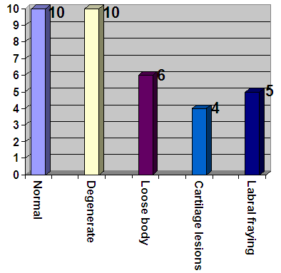
Figure 3. Predominant arthroscopic findings
No perineal or foot pressure sores were detected after surgery.
90% of our patients went home the same day. No
intra-operative or any major post-operative complications were
identified. (Table 1) Traction neuropraxia was observed in two
patients with paraesthesia in the leg and foot. Both were
transient and resolved completely. One patient with dysplastic
hip on whom diagnostic scope was performed had severe pain on
mobility in the post-operative period which delayed discharge.
Non-weight bearing for six weeks was the rehabilitation plan for
a hip with micro-fracture for isolated osteoarthritic lesion.
|
Age
/ Sex
|
Indication
|
Scope
findings
|
Complication
|
Outcome
|
|
35
F
L
hip pain
|
Acetabular
sub- chondral lesion
|
Degenerative
|
Paraesthesia
symptoms:
resolved by 3 months
|
Discharge
|
|
41
F
Dysplastic
R hip
|
Diagnostic
|
Degenerative
|
Poor
mobility-home in 3/7
|
awaiting
triple pelvic osteotomy
|
|
33
F
R
hip pain
|
Acetabular
degenerative changes
|
Labral
fraying
|
Paraesthesia
of foot: resolved same day
|
Discharge
|
Table 1: Complications
Of the study group, three patients did not turn up for their
initial follow-up visit. Three fourths of the rest (24 patients)
had a management plan at six weeks initial follow up and eight
patients needed to be reviewed for a longer period. Fourteen
patients were discharged with satisfactory outcome from our
clinic. Eighteen patients were referred to other specialists for
further treatment. Of these, data was available for sixteen
cases. Nine patients went on to be operated or are awaiting
surgical treatments like resurfacing arthroplasty, triple pelvic
osteotomy or femoral de-rotation osteotomy. Four were managed
non-operatively. One patient each was referred to the pain team
for ongoing hip pain, spinal surgeons for back pain and for
footwear modification for mild limb length inequality.
Discussion :
Hip
arthroscopy was first described by Burman in 1931 who had
stated: "It is manifestly impossible to insert a
needle between the head of the femur and the acetabulum".2
The ball and socket nature of the joint, its natural
intra-articular vacuum and surrounding neurovascular structures
make insertion of the arthroscope difficult and fraught with
danger. It was only in the 1980s that hip arthroscopy gained
recognition as a diagnostic and therapeutic procedure, to be
performed only by the experts.
It
cannot be overemphasized that proper patient selection is the
key to a successful outcome. Pathology confined to the hip joint
that is amenable to arthroscopic intervention and
reasonable expectations of postoperative outcomes are
the ideal selection criteria.
General
anesthesia or regional spinal anesthesia is equally effective
but adequate muscle relaxation is essential for joint
distraction. The patient can be positioned supine or lateral
decubitus position, the choice depending on surgeon preference.3,
4 We prefer supine approach for the simplicity of the
patient positioning, avoiding the need for specialized
distraction devices, familiar joint orientation and optimal
access for all portal placements.
Commonest
indications described in literature include diagnostic
arthroscopy, removal of loose bodies, synovial biopsy, subtotal
synovectomy, management of labral tears, synovial
chondromatosis, osteochondritis dissecans, chondral lesions, and
the treatment of septic arthritis.5 Contraindications
to hip arthroscopy include systemic illness, superficial
infection, arthrofibrosis or ankylosis,
non-progressing avascular necrosis and morbid obesity.
Patients aged above fifty five years and those with advanced
degenerative arthritis do not respond well to hip arthroscopy
and should be best avoided in them.6
Three
patients were in the paediatric age group (under 16 years) in
our study. Two boys had post traumatic loose bodies and one
underwent arthroscopic assessment of the articular surface. None
of the patients in our study were aged above 55 years .We did
not encounter inability to access the hip in any patient, though
this was found to be difficult in some.
|
Pre-operative diagnosis
|
Arthroscopy findings
|
|
Idiopathic painful hip(7)
|
Normal
|
|
|
Normal
|
|
|
Normal
|
|
|
Grade 3 and 4 degenerative changes
|
|
|
Acetabular grade 2 degenerative
changes
|
|
|
Labral fraying
|
|
|
Acetabular Chondral lesion
|
|
Labral pathology (6)
|
Normal
|
|
|
Normal
|
|
|
Normal
|
|
|
Grade 4 degenerative changes
|
|
|
Acetabular Chondral lesion
|
|
|
Extensive Labral tear
|
|
Sub-chondral (6)
|
Normal
|
|
|
Normal
|
|
|
Grade 4 degenerative changes of the
head of femur
|
|
|
Grade 4 degenerative lesion on the
femur head
|
|
|
Acetabular Chondral lesion
|
|
|
Acetabular grade 2 degenerative
changes
|
|
OCD
|
OCD lesion
|
|
Synovitis (nodular
thickening)
|
Labral fraying
|
|
Synovitis
|
Normal
|
|
Loose body (6)
|
Solitary osteochondral loose body
|
|
|
Solitary osteochondral loose body
|
|
|
Solitary osteochondral loose body
|
|
|
Solitary osteochondral loose body
|
|
|
Multiple osteochondral loose bodies
|
|
|
Multiple cartilaginous loose bodies
|
|
Diagnostic (4)
|
Normal
|
|
|
Chondral flap lesion
|
|
|
Gr. 4 degenerative changes of fovea,
Gr. 2,3 rest of hip
|
|
|
Grade 2 and3 degenerative changes
superior acetabulum
|
|
OA (3)
|
Grade 4 degenerative changes of the
head of femur
|
|
|
Grade 4 degenerative changes of the
head and acetabulum
|
|
|
Posterior labral fraying
|
Table 2. Pre-operative diagnosis and post operative
findings
Overall
arthroscopy altered the diagnosis in all but 11 patients. (Table
2) This did not include the four hips in which hip scope was
performed to asses the articular surfaces. Even in this group,
arthroscopy revealed degenerative changes in two hips and
cartilaginous lesion in one hip. Of the seven patients with a
working diagnosis of idiopathic painful hip, arthroscopy altered
the diagnosis in four hips. The new diagnosis included
osteoarthritis in 2, osteochondral lesion in 1 and labral
fraying in one. Previous studies have shown the usefulness of
diagnostic arthroscopy compared to MRI in reliably detecting
chondral lesions and cartilaginous loose bodies.7, 8
In
our study, a correlation between MRI result and arthroscopic
finding regarding labral tear was seen in only one hip. Five
patients with a labral lesion shown by imaging studies did not
demonstrate the lesion on arthroscopy. One hip had labral
fraying not detected by MRI. As Dorfmann et al have shown in
their study, labral lesions were commonly overestimated at
arthrography and only 18 lesions of 413 hips (4.4%) were
confirmed on arthroscopy.9
In
the 24 hips with a preoperative diagnosis, arthroscopy revealed
a different finding in 15 (62%). Of these, six patients had
normal looking hips. The others with different diagnosis include
osteoarthritis in 5, labral fraying in 2 and one osteochondral
lsion. One hip with
preoperative imaging suggesting intra-articular osteochondroma
showed multiple cartilaginous loose bodies, an outcome of
advanced osteoarthritis.
Our
study showed two nerve related complications with patients
complaining of paraesthesia in the sciatic nerve distribution.
Both of these were transient and made complete recovery. One
patient had difficulty in mobilization during rehabilitation
period and was discharged home on the third post-operative day.
The reported complication rate in literature is between 0.5 to 5
%.11 The commonest are traction neurapraxia, direct
trauma to neurovascular structures, pressure sores, and
largely unreported, iatrogenic joint damage. Rarer described
complications include myositis ossificans, fluid extravasation,
reflex sympathetic dystrophy, trochanteric bursitis, labial
injury and instrument failure.
Post-operative
rehabilitation protocols following hip arthroscopy is only
recently coming into limelight. Rehabilitation protocols that
have been typically used for surgeries such as total hip
arthroplasty are often not sufficient for the population of
patients undergoing arthroscopic procedures of the hip joint.12
Postoperative rehabilitation can be staged into three phases
under the supervision of the physical therapist. The initial
phase in the first few weeks aims at restoring range of
movements within tolerance and progressing on to full weight
bearing. Weight bearing may be limited after some surgical
procedures with a hip arthroscopy, including labral repair,
iIliopsoas release, microfracture and capsulorraphy. The next
few weeks comprise the intermediate phase, where the goal is to
regain and build muscle strength. Finally the advanced phase
involves improving the functional strength, endurance and
stability with gradual return to sporting level activity as
necessary.
Outcome
measure studies have shown favourable results from hip
arthroscopy in selected indications. OLeary et al from 86
hips have shown best results in patients with labral injury,
late Perthes disease, loose bodies or focal chondral defects
and poor results in avasular necrosis and degenerative
arthritis.13 They conclude that the presence of
mechanical symptoms is a favorable prognostic factor for any
diagnosis except degenerative arthritis. Byrd et al in their
prospective analysis of 121 cases, have identified that patients
with acute or traumatic onset of symptoms with greater
improvement than those with insidious onset of symptoms and that
longer duration of symptoms especially in male counterparts
correlated with less successful outcomes.14 Greatest
symptomatic improvement was noted in arthroscopic removal of
loose bodies. The authors opine that hip arthroscopy can be
performed for a variety of conditions (except end-stage
avascular necrosis) with reasonable expectations of success.
Baber et al have showed that arthroscopy revealed an abnormality
in 81% of idiopathic painful hips and found a different
abnormality in 30% of patients with a preoperative diagnosis.10
They report that arthroscopy aided in management in
seventy four percentage of hips either by a change in the
primary diagnosis in (53%) or by improvement of symptoms( 21%)
Their study advocates the role of diagnostic arthroscopy
especially with early cartilaginous lesions, labral tears and
loose bodies.
Conclusion:
The
role of hip arthroscopy in the management of hip disorders
continues to expand with continued experience and improved
instrumentation. It is now becoming increasing used for surgery
to the structures surrounding the hip, not just to those within
the hip cavity. Whatever the method, the most critical
determinants for a successful outcome are patient selection and
surgical expertise. Patients with mechanical symptoms and
pathology confined to the hip joint and a reasonable expectation
of the outcome are the ideal candidates for hip arthroscopy.
Awareness of the potential complications, attention to patient
positioning and proper orientation of portal sites is the
surgeon factor that dictates good outcome.
Our
study in the small, heterogenous group of patients with hip pain
has shown hip arthroscopy to be a safe and effective means for
assisting the management of hip disorders.
Reference :
-
Diulus
CA, Krebs VE, Hanna G, Barsoum WK.
Hip arthroscopy technique and indications. The Journal of
Arthroplasty. 2006 June; 21(4 Suppl 1):68-73.
-
Burman MS. Arthroscopy or the direct visualisation of joints: an
experimental cadaver study. Journal of Bone & Joint Surgery
1931; 8:669-95.
-
Glick
JM, Sampson TG, Gordon RB, Behr JT, Schmidt E. Hip
arthroscopy by the lateral approach. Arthroscopy. 1987;
3(1):4-12.
-
Byrd
JW. Hip arthroscopy utilizing the supine position.
Arthroscopy. 1994 June; 10(3):275-80.
-
McCarthy JC, Busconi B. The role of hip arthroscopy in the
diagnosis and treatment of hip disease. Orthopedics. 1995;
18:753-756.
-
Carreira
D, Bush-Joseph CA. Hip arthroscopy. Orthopedics. 2006 Jun; 29(6):517-23.
-
PalmerWE.
MR. Arthrography of the Hip. Seminar in Musculoskeletal
Radiology. 1998; 2(4):349-362.
-
Edwards
DJ, Lomas D, Villar RN. Diagnosis of the painful hip
by magnetic resonance imaging and arthroscopy. Journal of Bone
& Joint Surgery
Br.
1995 May; 77(3):374-6.
-
Dorfmann H, Boyer T. Arthroscopy of the Hip: 12 Years of Experience. Arthroscopy. Vol. 15, No. 1,
1999, 67-72.
-
Baber
YF, Robinson AH, Villar RN. Is diagnostic arthroscopy
of the hip worthwhile? A prospective review of 328 adults
investigated for hip pain.Journal of Bone & Joint Surgery
Br. 1999 Jul; 81(4):600-3.
-
Clarke
MT
, Arora A, Villar RN. Hip arthroscopy: Complications in 1054
cases. Clinical Orthopaedics and Related Research .2003 Jan;
406: 84-88.
-
Enseki
KR, Martin RL, Draovitch P, Kelly BT, Philippon MJ, Schenker ML.
The hip joint: arthroscopic procedures and postoperative
rehabilitation. Journal of Orthopaedic and Sports Physical
Therapy2006 Jul; 36(7):516-25
-
O'leary
JA, Berend K, Vail TP. The relationship between diagnosis and outcome in
arthroscopy of the hip. Arthroscopy. 2001 Feb; 17(2):181-8.
-
Byrd
JW, Jones KS. Prospective analysis of hip arthroscopy
with 2-year follow-up. Arthroscopy. 2000 Sep; 16(6):578-87.
|

