| CASE
REPORT |
|
Asymptomatic
Traumatic Posterior Dislocation Of Sternoclavicular Joint In A
Child: A Case Report
|
|
Singh
VK*,
Singh PK*, Mishra
A*, Tomar J*
*
James
Paget
University
Hospital
, Great
Yarmouth
,
UK
Address for Correspondence:
Mr. V.K Singh, MRCS.
Department of Trauma & Orthopaedics Surgery,
James Paget University Hospital, Great Yarmouth, U.K. NR31 6LA
Email : we.publish@googlemail.com
|
|
Abstract:
Injury to
Sternoclavicular joint is uncommon but may be a life threatening
if the diagnosis is not made acutely. Posterior sternoclavicular
joint dislocation is associated with a number of complications
including trachea tear
or trauma to the great vessels. Diagnosis by conventional
radiography is difficult. Even experienced examiners may miss
the diagnosis unless a high level of suspicion exists and the
appropriate imaging studies are ordered. We report a case of
traumatic dislocation of Seternoclavicular joint in an
11-year-old child post rugby injury, successfully treated by
closed reduction.
J.Orthopaedics 2007;4(3)e14
Keywords:
Sternoclavicular joint; bilateral; dislocation; child; sports
injury.
Introduction:
Sternoclavicular
joint (SCJ) is a synovial diarthrodial joint that links upper
extremity to Torso. It participates in all the movements of
upper extremity by providing free movement of clavicle in
virtually all directions. Stability of this joint is imperative
for ability to thrust the arm and shoulder forwards. Articular
surface of clavicle is much larger than articular surface of
sternum hence only 50% of the medial end of the
clavicle articulates with sternum. SCJ has little inherent
stability. Most of the strength and stability originates from
the joint capsule and supporting ligaments. The capsule
surrounding the joint is weakest inferiorly, while it is
reinforced on the superior, anterior, and posterior aspects by
the various ligaments. These include the interclavicular,
anterior and posterior sternoclavicular and costoclavicular
ligaments. Sternoclavicular joint dislocations are rare
injuries1, 2 accounting for <1% of all traumatic joint
dislocations. It was first described in 1824 by Cooper3 and
since then only around 100 cases of posterior dislocation of SCJ
have been reported in literature. Anterior dislocation is more
common4 than posterior dislocation (9:1). Most of these injuries
have been described in adults. We report a case of asymptomatic
traumatic posterior dislocation of sternoclavicular joint in an
11year old child post sporting injury.
Case Report :
An 11-year-old enthusiastic rugby player was injured while playing rugby. He was involved in a bad tackle and fell awkwardly on his right shoulder and another player fell on top of his chest. He had to discontinue playing because of pain. He was assessed by the physical trainer who advised him to visit casualty for assessment.
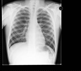
In casualty child was comfortable with minimal pain and had restricted range of right upper limb movements. He had swelling, minimal tenderness and a subtle palpable dip in right sternoclavicular area. He had no other symptoms and was neurovascularly intact. Child was sent for a chest X-ray to look for possible sternoclavicular dislocation, which was inconclusive (Fig 1).
|
 |
 |
|
Fig.1 The X-ray, taken in A&E, immediately after admission. The radiograph is unable to reveal any abnormality and was reported normal by radiologist. |
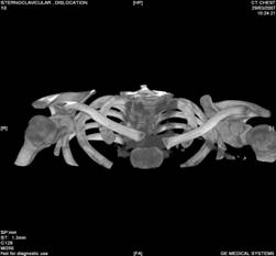
Fig 2: The high
resolution CT scan with 3- dimensional reconstruction of
thoracic cage. The scan reveals the separation of both
clavicles from their sternal attachments with posterior
displacement (Arrow Heads)
|
|
Specialist opinion was sought because of clinical suspicion. Child was admitted under the care of orthopaedic team for observation and CT scan to rule out dislocation. There was no
change in patients symptoms and CT scan was done next day, which revealed posterior
dislocation of both sternoclavicular joints (Fig 2). Once the diagnosis is confirmed he
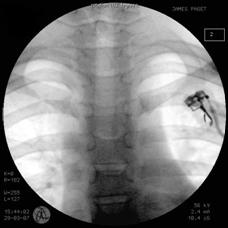
was urgently taken to theatre. Successful closed reduction was performed under general anaesthetic using fluoroscopic guidance (Fig 3).
His symptom resolved post manipulation and was subsequently discharged home. Follow up in clinic in showed normal
examination and complete resolution of symptoms.
|
 |
|
Fig 3: The intra-operative image showing the satisfactory |
|
Discussion :
Injuries to the sternoclavicular joint are uncommon, but can be life-threatening if not diagnosed and treated adequately. Nearly 25% of patient with SCJ dislocation have associated injuries5 such as pneumothorax, injury to brachial plexus, laceration of the superior vena cava, occlusion of the subclavian artery or vein, and disruption of the trachea. All of these complications can cause significant disability and even death. Prompt diagnosis and treatment is necessary to avoid complications.
High index of suspicion in needed in case of high velocity road traffic injuries or high impact sporting injuries. Children playing contact sports are particularly at risk. Chest X-ray may help in diagnosis but should not be used to rule out SCJ dislocation. Specific view of SCJ dislocation such as Rockwood (serendipity), Hobb, Kattan6 and Heinig views may be requested to help diagnosis but CT scan is the imaging modality of choice. It is imperative to make the diagnosis early, as delay may be life threatening, and closed reduction is best accomplished during the first 48 hours.
When diagnosis is not clear patient should be admitted for adjunctive imaging (CT scan) to rule out the diagnosis. Reduction of posterior dislocation must only be done in theatres under general anesthetic7 in view of the possibility of compression of mediastinal structures8 with cardiothoracic team on standby.
Reference :
|
-
Cope R, Riddervold HO, Shore JL, Sistrom CL. Dislocation of the sternoclavicular joint: anatomic basis, etiologies, and radiologic diagnosis Orthop Trauma1991; 5:379384.
-
Denham RH, Jr., and Dingley AF, Jr.: Epiphyseal Separation of the medial end of the clavicle.J.Bone And Joint Surg., 49-A: 1179-1183, Sept.1967.
-
Cooper A: A Treatise on Dislocations and on Fractures of the Joints, Ed 3, p 559. London, Longman, 1824.
-
Rockwood CA, Wirth MA. Injuries to the sternoclavicular joints. In: Rockwood CA, Green DP, Bucholtz RW, Heckman JD, eds. Fractures in adults, 4thed. Philadelphia: Lippincott-Raven, 1996:14151471.
-
Atraumatic posterior dislocation of the sternoclavicular joint, Clin Orthop Relat Res.1993 Jul;(292): 159-64.
-
Kattan KR. Modified view for use in roentgen examination of the sternoclavicular joints Radiology.1973; 108:8.
-
Selesnick FH, Jablon M, Frank C, Post M. Retrosternal dislocation of the clavicle: report of four cases. J Bone Joint Surg. 1984; 66A: 287291.
-
Sanders JO, Lyons FA, Rockwood CA. Management of dislocations of both ends of the clavicle. J Bone Joint Surg. 1990; 72A: 399402.
|
|
This is a peer reviewed paper Please cite as
: Singh
VK : Asymptomatic
Traumatic Posterior Dislocation Of Sternoclavicular Joint In A
Child: A Case Report
J.Orthopaedics 2007;4(3)e14
URL:
http://www.jortho.org/2007/4/3/e14 |
|
|

