|
Abstract:
A
review of 110 cases of intertrochanteric fractures
treated by using Dynamic Hip Screw /Dynamic Martin Screws over a
period of two years was done. Medial bony contact was obtained
before fixation of the fractures. In cases with doubtful medial
cortical stability autogenous bone grafting from iliac crest was
done. Excellent to good results were obtained in 72 of the cases
(66.4%). 32 cases (29.1%) showed satisfactory results. 6 cases
(5.5%) had shown poor results.
Keywords: Intertrochanteric fractures, Dynamic Hip Screw, Autogenous
bone grafting.
J.Orthopaedics 2007;4(2)e40
Introduction:
Intertrochanteric
fractures have been estimated to occur in over 2,00,000 patients
in the United States (1). Reported mortality with these
fractures ranges from 15 -20%. In its natural history these
fractures are known to occur in elderly patients. . These
fractures are thrice more frequent in women than in men. Due to
increase in average life expectancy as
a result of better medical care our society is having a
large number of aged population those are more prone to
intertrochanteric fractures. Surgical management is the
treatment of choice for these fractures as to mobilize the
patients out of the bed and thus preventing complications of
prolonged recumbency. In this paper we review our experiences
with Dynamic Hip Screw/Dynamic Martin Screw in the management of
intertrochanteric
fractures.
Material and Methods :
A total of 110 cases managed at three Service Hospitals were
reviewed in this study.
A thorough clinical and radiological evaluation was done for
every patient. Fractures were classified as per A. O. Group
classification for intertrochanteric fractures (2).
Patients were taken up for surgery within 7 -15 days.
Preoperative skeletal traction immobilization was done in the
ward. Surgery was
performed mostly under spinal anesthesia and occasionally under
general anesthesia depending upon the general condition of the
patients. Patients were positioned supine on Albees fracture
table. Trochanteric region was exposed via lateral approach and
guide wire was passed under image intensifier guidance. Fixation
was done by using DHS/ DMS.
Unstable fractures geometry was made stable by obtaining medial
cortical bone contact between femoral neck, trochanter and upper
shaft. Post operatively, drain was removed after 48 hours and
patients were encouraged to move out of the bed. Non - weight
walking was started by 5th day. Suture removal was done after 14
days and patients were discharged home with the advice to walk
non-wt bearing with crutch/walker support for a period of 4 to
6-wks depending on the comminution at the fracture site. Partial
weight bearing was started thereafter till the consolidation of
fracture fragments. Follow up was done up to a period of 2 yrs.
Results :
A total of 110 hips were
operated during this period. No of male patients were71 (64.5 %)
and the females were 39 ( 35.5 %) . All the patients had
unilateral intertrochanteric
fractures. The age distribution was as under.
Table1
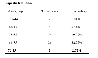
The youngest patient in our series was 35
years old and the eldest was 78 yeas old. Maximum number of
cases was in the age group range of
56-65 years. All
the patients
Sustained
direct trauma to the hip having usually involved in a road
traffic accidents or accidental falls. All the
patients were operated under spinal anesthesia. All the cases in
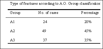
our series were operated. According to AO classification the
distribution was as under.
Table
2
Patient not operated due to various reasons were not
considered in
this study. There was no mortality in our series. Patients were
ambulant non-weight bearing after 5th post- operative day using
a walker. Isometric exercises were started after 48 hours.
Antibiotics were used for 5 days with first dose given at time
of incision. Post- operative infection occurred in two cases.
First case had superficial wound infection and second
case had delayed wound infection, six months after the
surgery. These cases were managed
with debridement and antibiotics.
Discussion :
In
our series 110 cases of intertrochanteric fractures were managed
with Dynamic Hip Screw /Dynamic Martin screw fixation. Patients
managed conservatively due to various medical & other
related problems were not included. This was done to observed
the natural history of this treatment of modality .The male:
female ratio in our series is 1.8:1
Average age in these patient is 55 to 75 years .In
unstable fractures medial bone contact was
obtained before fixation of the fracture .
As demonstrated by Wolfgang (3) approximately 10%) medial
movement of unstable fracture resulted in 90% of union. The use
of sliding device theoretically allowed & unstable fracture
to impact and there by seek its own position of stability. Jacob
& colleagues(4) demonstrated that as
the sliding device shortens with settling of an unstable
fractures, the lever arm acting on the nail plate junction
shortens thereby reducing the force on the implant. Clowson(5),
Ecker and colleagues(6) noted that unstable fractures
treated with sliding device
underwent shortening and medial displacement but fracture
went on to prompt union. Although
shortening upto
1 cm occurred the head did not fall into
varus, nor did the fixation device cut through the head
and damaged the acetabulum
. In our series we had obtained medial and posterior bony
stability in treating the unstable fracture and if doubtful bony
contact was noticed, autogenous bone grafting from ipsilateral
iliac crest was done. We did not have any cases of implant
failure and proximal migration of implant into the joint. As
noted by Wolfgang and coworkers (3)
that unstable fracture treated without obtaining bony
stability had 21 % mechanical failure. This rate was reduced to
10% when bony stability was obtained.
Jenson(7) and Moore(8) reported a 10% in hospital
mortality rate associated with
intertrochanteric fractures. In our series fortunately we
had no in hospital mortality. The incidence of post-operative
wound infection after open reduction and internal fixation of
intertrochanteric fracture varies from 1.7% to 16.9%(9,10 ). In
our series we had two cases (Case No.ll and 23) of wound
infection (1.8%). One case has superficial wound infection
(group 1 as per Barr's(11)four group of
post operated infection). Second case had group III (late
sepsis after six months). These cases were managed with local
wound debridement and wound dressing. Intertrochanteric fracture
occur in cancellous bone with good blood supply, non
union has been found to be uncommon. The incidence of non-union
is reported to be 1 % -2%(12). Those
intertrochanteric fractures prone to non union includes
comminuted unstable
fracture with loss of medial calcar continuity, which when
stabiIised tends
to fall into varus.
Mariani and Rand(13) reported that 10 of 20 patients with non-
union of intertrochanteric fractures has unstable fractures with
loss of medial support. Laskin
and associates(14) noted that union was present within
three months of fracture and that non union was secondary to
poor bony opposition at the time of surgery. In our series we
did autogenous bone grafting having doubtful medial bony support
thus there were no non union noted in our series . We
started partial weight bearing after 4 weeks in patients whom
stable internal fixation was obtained .Patients having unstable
fracture geometry were allowed partial weight bearing after 8-12
weeks. Full weight bearing was allowed after complete
radiological union for all the cases on individual basis.72
patients had good function recovery and sound bony union.32
patient had satisfactory results with shortening up to2.5
centimeters and few degree of terminal restriction of hip joint
movements. 6 cases has shown poor results with shortening of
more than 3 centimeters and varus angulation of 100
degrees. These patients had painful restriction of hip joint
movement and limp while walking.In our review we concluded that
sliding compression hip screw the implant of choice
for the treatment of intertrochanteric fractures. We also
recommend primary bone grafting from ipsilateral iliac crest in
unstable fractures where medial bony contact is poor.
Reference :
-
James L.Guyton. In: Campbells operative orthopaedics.
S.Terry Canale, M.D.editor. 9th ed. 1998: 2182-2183, Mosby
Year Book, Inc, USA.
-
Muller ME, Allgower M, Schneider R, Willenegger H; Manual
of Internal fixation: techniques recommended by the AO, ASIF
group, ed 3, Berlin, 1991 Springer-Verlag.
-
Wolfgang, G.L.; Bryant, M.H. ; and ONeill, J.P.;
Treatment of Intertrochanteric Fracture of the Femur Using
Sliding Screw Plate Fixation Clin.Orthop., 163:148-158,1982
-
Jacobs, R.R; McClain, O.; and Armstrong, H. J.; Internal
Fixation of Intertochanteric Hip Fractures: A Clinical and
Biomechanical Study. Clin. Orthop., 146:62-70,1980.
-
Clawson, D.k.; Trochanteric Fractures Treated by the
Sliding Screw Plate Fixation Method.J.Trauma, 4:737-756,1964.
-
Ecker, M.L.; Joyce, J.J.; and Kohl, E.J; The Treatment of
Trochanteric Hip Fractures Using a Compression Screw. J. Bone
Joint Surg., 56A23-27, 1975.
-
Jensen, J.S. : Trochanteric Fractures. Acta Orthop.
Scand. (Suppl.), 188:1-100,1981.
-
Moore, M. ; Treatment of Trochanteric Femoral Fractures
With Special Reference to Complications. Am. J. Surg.,
84:449-452, 1952.
-
Cleveland, M, Bosworth, D.M, and Thompson, F.R.;
Intertrochanteric Fractures of the Femur. J. Bone Joint Surg.,
29:1049-1067,1947.
-
Kyle, R.F.; Intertrochanteric Fractures. In Chapman M.W.
(ed.) : Operative Orthopaedics,pp. 353-359. Philadelphia,
J.B.Lippinoctt,1988.
-
Barr, J.S.; Diagnosis and Treatment of Infections
Following Internal Fixation of Hip Fractures. Orthop. Clin.North
Am. 5:847-864,1974.
-
Wilson,H.J.:Rubin,B.D.;Helbig;F.E.J.;Fielding,J.W.;and
Unis,G.L.; Treatment of Intertrochantric Fractures with Jewett Nail: experience with 1,015 cases.Clinical
orthopaedics .,148:186-191,1980.
-
Mariani,E.M.,and Rand,J.A.;Subcapital Fractures after
open reduction and internal fixation of Intertrochantric
Fractures of the Hip . Clin Orthop, 245:165-168,1989.
-
Laskin,R.S.:Gruber,M.A.and Zimmerman,A.J.;
Intertrochanteric Fractures of the Hip in Elderly :A
Retrospective Analysis of 236 cases . Clin Orthop.141:
188-195,1979.
|

