| CASE
REPORT |
|
Deformed
Tibial Nails: A Case Report and Biomechanical analysis
|
|
Mohan
Pullagura*, Paul Banaszkiewicz
*Department
of Trauma and Orthopaedic Surgery
Queen Elizabeth Hospital Sheriff Hill,
Gateshead,
UK
Address for Correspondence
Paul
Banaszkiewicz, Dept of Orthopaedics,
Queen Elizabeth Hospital,
Sheriff Hill, Gateshead, Newcastle. UK. NE9 6SX
E-mail: mohanpk73@yahoo.co.in
pbanaszkiewicz@hotmail.com
Tel:
00447766832733, 00441914453214
|
|
J.Orthopaedics 2007;4(2)e4
Introduction:
Intramedullary
nailing (IM) is regarded as the procedure of choice for
stabilization of displaced diaphyseal fractures of the tibia[3].
This is a load-sharing device, which allows early weight bearing
through bone.
There
are several reported reasons to remove IM nails. These include
sepsis, pain and irritation at the point of insertion, the
difficulty of treating a new fracture if a nail is in place
because the nail can be deformed or the fracture can occur
around or at the end of the nail (new fractures are much more
difficult to manage when a nail is in place) and breakage of
nails following fracture healing.
Excessive
loading or repetitive cyclic loading can cause fatigue fracture
of a nail especially if the fracture has not fully healed[2].
A high-energy injury can lead to deformation and angulation of
the nail. Biomechanically the weakest point is at the site of
fracture healing, hence the most common site of angulation[1].
Although
it is uncommon to sustain a second high-energy injury after a
recent nailing, some patients are eager to return to sports as
quickly as possible, which increases the risk of subsequent
trauma[11].
The deformation depends on the tensile strength of the nail
(diameter), weight of the patient, time of original injury
(fracture healing status) and the forces going through the nail
and tissue envelope[6].
Case Report :
A
32-year-old man involved in a road traffic accident sustained a
closed fracture of the middle thirds of his right tibia. The
patient was treated with primary IM nailing on the same day. The
medullary canal was reamed to 11mm and a 10mm Î
380mm cannulated ST-Pro tibial nail (Grosse-Kempf model)
inserted with 2 locking screws proximally and distally.
Postoperative radiographs demonstrated that normal tibial length
was restored and the alignment of the fractured bone was normal
in all planes. Post-operatively he made an uneventful recovery
and was allowed to bear full weight within 4 weeks. After seven
months the locking screws were removed under local anaesthesia
because they were backing out. The fracture had radiographically
united at this stage with callus formation seen on all four-bone
cortices in AP and lateral radiographs. Eight months following
IM nailing he sustained a second injury when two 15 stone
players fell on him whilst playing football. The plain
radiographs showed an angulation at the original fracture site
and a deformed nail at the same level. The angulation was 11
degree valgus and 26 degree posterior. He was taken back to the
theatre and attempted closed in-situ straightening of the nail
performed using the three-point fixation principle. The bent
nail could only be partially straightened but despite this the
nail was successfully removed. Unfortunately it was reluctantly
accepted that unavoidable fracture extension would occur with
nail removal. The canal was over reamed to 12mm with particular
attention to remove fibrous tissue around the fracture site and
a 11mm Î
380mm IM nail inserted. The postoperative recovery was
uneventful and solid radiological union was noted at 2 months.
|
|
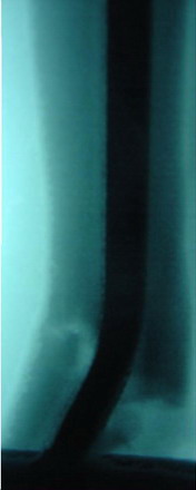
Fig.1 Antero-posterior view plain radiograph showing deformed
nail
|

Fig 2 Antero-posterior
view plain radiograph showing healing after re-nailing
|
|
Discussion :
There
is good evidence that fracture stiffness is a reliable predictor
of healing[10],
to allow safe return to independent weight bearing and sedentary
activities[8].
IM nails have no function after fracture healing[5].
Nails and interlocking screws tend to fail by plastic
deformation or fatigue fractures and the failure rate is
reported to be about 5-10%[4].
However routine removal of nails is not mandatory because the
refracture can be as high as 21% depending on the implant design[9].
There are a few authors who advocate removal of nails 1-2 years
after the injury in young and vigorous patients[5].
Even
in the presence of osseous bridging across the fracture
fragments and with the nail sharing the load of the weight an
excessive stress applied has lead to deformation of the nail and
refracture through the bone. It has been shown that it takes
eighteen months for bone to regain its tensile strength to 80%
of the original. Good patient compliance is needed for the
osseous healing process of the extremity and conscious exertion
of stress (weight bearing) is just as important for healing as
an implant material, which should withstand the unexpected
excessive stress in the early phases of healing[7].
The
implant diameter has been definitely shown to influence the
bending strength by a factor of 4. However a 10mm or 11mm nail
or a healed fracture cannot achieve the stiffness of the intact
tibia.
The
biomechanical analysis of the nail showed no corrosion or wear
in the metal. The 10mm cannulated nail had a stiffness of 64.3
± 6.1 Nm² and a yield bending moment of 86.2 ± 3.7 Nm[8].
The force required to permanently bend a nail of this diameter
without any contribution from the bone-to-bone contact or from
the foot contact with the floor was 718 N (body weight of a 73
kg person). Assuming the bone is healed with the foot on ground
covered with the whole soft tissue envelope it would require 10
times the force to cause the same deformation. Therefore to
cause re-fracture and nail deformation a force of between
718N(body weight of a 73 kg person) to 7180N(body weight of 730
kg) was required.
Conclusion:
In
our case the patient went back to active sports after eight
months with good clinical and radiological signs of union. The
tensile strength of bone was not fully re established. We
would like to conclude that it might be better to wait till
eighteen months before allowing patients to get back to active
contact sports or high speed motor diving. This will avoid the
complications of refracture, more soft tissue injury or nail
fracture while attempting to straighten out the nail or
extraction of the nail.
Reference :
-
Apivatthakakul
T, Chiewchantanakit S: Percutaneous removal of a bent
intramedullary nail. Injury. Nov; 2001; 32(9): 725-6
-
Burzyski N,
Scheid DK: A modified technique for removing a bent
intramedullary nail minimizing bone and soft tissue
dissection. Journal of Orthopaedic Trauma 1994; 8:181-182
-
Court-Brown
CM, Will E, Christie J, McQueen MM. Reamed or Unreamed
nailing for closed tibial fractures. J Bone and Joint
Surgery 1996; 78B: 580-583,
-
Cunnigham J.
The Biomechanics of Fracture Fixation. Current Orthopaedics
2002; 457-464
-
David
Seligson, Howard P A, Martin R. Difficulty in Removal of
Certain Intramedullary Nails. Clinical Orthopaedics and
Related Research 1997; 340,p202-206
-
Kelsch G,
Kelsch R, Ulrich C. Unreamed tibial nail (UTN) bending: case
report and problem solution. Arch Orthop Trauma Surg 2000;
123:558-562, 2003
-
Reeves EA.
Testing of ST-Pro tibial nails to ASTM F1264-96b. Biomet
Merck Ltd. R&D 2005; Test Report 72
-
Richardson J
B, Cinningham J L, Goodship A E, OConnor B T, Kenwright
J. Measuring stiffness can define healing of tibial
fractures. Journal of Bone and Joint Surgery (Br); 1994;
76:389-394
-
Takakuwa M,
Funakoshi M, Ishizaki K, Aono T, Hamaguchi H. Fracture on
Removal of the ACE Tibial Nail. Journal of Bone and Joint
Surgery (Br) 1997, 79,3, p444-445
-
Wade R H,
Moorcroft C I, Thomas P B M. Fracture stiffness as a guide
to the management of tibial fractures. Journal of Bone and
Joint Surgery (Br); 2001, 83,4:533-535
-
Yip KMH,
Leung KS. Treatment of Deformed Tibial Intramedullary nail:
Report of Two Cases. J Orthop Trauma 1996; 10:580-583
|
|
This is a peer reviewed paper Please cite as
:Mohan
Pullagura :Deformed
Tibial Nails - A Case Report and Biomechanical analysis
J.Orthopaedics 2007;4(2)e4
URL:
http://www.jortho.org/2007/4/2/e4 |
|
|

