|
J.Orthopaedics 2007;4(2)e1
Introduction:
Scheuermann's Disease (Osteochondritis of
vertebral epiphyseal plates or Adolescent kyphosis) is one of
the most confusing and poorly understood abnormalities of the
adolescent spine. It consist of primary irregularity of
ossification of one or more vertebral end plates and leads to
thoracic kyphosis as name suggests [1,2]. We present here
early MRI findings in 2 patients of Scheuermanns Disease
before any kyphotic deformity.
Case Report :
Case 1: An 18 years
old lady presented with backache in the area of lower dorsal
spine for last 6 months. The pain increased on exertion and in
bending posture. There was no radiation of pain in the lower
limbs and no history of any neurological deficit. There
was no history of fever, trauma or any prolonged illness.
On
examination mild tenderness on palpation was present at D12
level. No swelling was seen in the back. . On neurological
examination, no abnormality or neurological deficiency was seen.
All relevant investigations including hemoglobin, ESR were
normal. Chest radiograph was normal.
Radiographs of the dorsolumbar spine were done in AP and lateral
projections. There was evidence of mild anterior wedging of the
D11 vertebra. End plates at D11, D12 levels were showing
thickening and sclerotic changes with mild concavity, more at
superior end plates of D11 and D12. Disc spaces at D10-11,
D11-12 levels were mildly reduced.
Then
MRI of the dorsal spine was performed on 0.2T GE signa MRI. T1W,
T2W sequences were taken in axial and sagittal planes with T2W
coronal and STIR sagittal sequences. On MRI, there was slight
anterior wedging of the D11 vertebra. Superior end plates of D11
and D12 vertebrae were showing irregularity and grade 1 to grade
3 degenerative changes with grade 3 changes predominating seen
as hypointensity on T1W, T2W, and STIR sequences.
Intervertebral discs at D10-D11 and D11-12 levels were showing
degenerative changes with loss of disc height and slight loss of
T2W hyperintensity because of dehydration (Fig. 1). Disc
material was herniating into the degenerated end
plates forming Schmorls nodes seen more clearly on T1W
sequence (Fig- 2). The degenerated discs were also
herniating anteriorly underneath anterior longitudinal ligament
(Fig. 1). Rest of vertebrae and disc spaces were normal.
Spinal canal was normal, spinal cord was normal. No pre or
paravertebral collection seen.
 |
 |
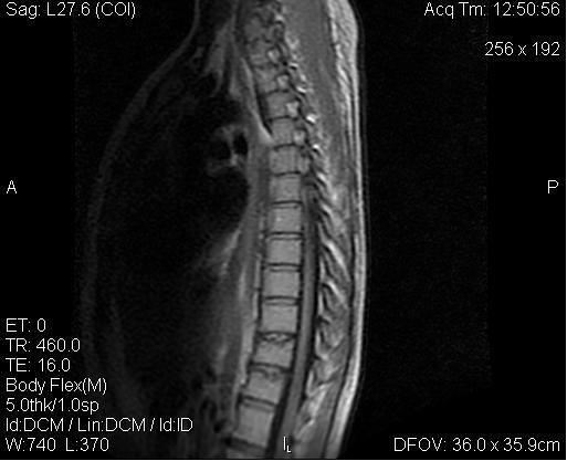
| Fig .1: Sagittal T2W image showing
degenerated D10-11, D11-12 intervertebral discs with end
plate sclerosis and anterior disc herniations |
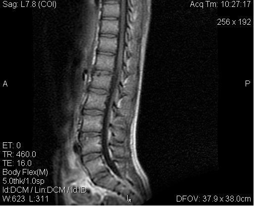
Fig. 2: Sagittal T1W sequence
showing anterior wedging of D11 with herniated disc
material at D11, D12 level forming schmorls nodes |
In adolescent patients
these findings of vertebrae wedging with end plate changes,
schmorls node formation and disc degeneration and bulges are
suggestive of a diagnosis of Scheuermanns
Disease.
Case 2: A 16 years old man
patient was presented with pain in back in lower dorsal and
lumbar area for last 1 year without any radiation to lower
limbs. There was no history of trauma, fever, neurological
deficit. On clinical examination neither swelling nor tenderness
was present. All blood investigations were normal. Radiograph of
the chest was also normal.
Radiographs of dorsolumbar spine were done in AP and lateral
projections. There was evidence of end plate sclerosis and
reduced D12-L1 disc space with small part of superior end plate
apophysis seen projecting anteriorly forming limbus vertebra.
Small schmorls node was also seen at superior end plate of L4
vertebra.
MRI of the dorsolumbar spine was done on 0.2T GE signa MRI. T1W,
T2W sequences were taken in axial and sagittal planes along with
STIR sequence in coronal and sagittal planes. There was
irregularity and sclerosis of superior end plate of L1 vertebra
and herniation of descicated D12-L1 disc material beneath the
degenerated end plate in anterior part of L1 vertebra (Fig-3,4)
suggestive of osteochondritis of L1 superior end plate
with herniation of descicated disc material. Degenerative end
plate changes with schmorls node formation because of disc
herniation were seen at superior end plate of L4 vertebra
(Fig-4). Spinal canal was normal. Spinal cord, thecal sac and
cauda equina were normal. Rest of vertebrae and discs were
normal. No evidence of bone marrow inflammation. No
abscess/collection seen in pre and paravertebral area. All these
findings in adolescent male patients are suggestive of early
Scheuermanns Disease.
 |
 |
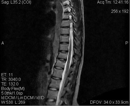
| Fig. 3:
Sagittal T1W image showing end plate sclerosis at L1
level and schmorls node with end plate degeneration
at L4 level.
|
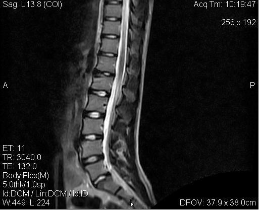
Fig. 4: Sagittal T2W image showing
descicated D12-L1 intervertebral disc herniating beneath
the degenerated superior end plate of L1 vertebra
forming limbus vertebra.
|
Discussion :
Scheuermanns Disease affects
mainly the dorsal spine of teenagers. This condition affects
both sexes .It usually begins at puberty with peak incidence
from 13 to 17 years. The mid and lower thoracic spine is the
region most commonly affected and usually several adjacent
vertebrae are involved. Less frequently the lesion may be found
in lumbar spine as was seen in one of our patients or upper
thoracic spine. Sometimes the changes are confined to single
vertebra [3].
Current criteria frequently require the presence of
abnormalities in at least three contiguous vertebrae, each with
wedging of 5 degrees or more. Such criteria however exclude
cases of Scheuermanns Disease that are associated with
vertebral irregularity without wedging as was seen in our
patients [4].
In some persons the disease is totally asymptomatic and in
others prominent symptoms and signs can be seen. Fatigue,
defective posture, aching pain affected by physical exertion and
tenderness to palpation are encountered mainly in midthoracic
and lumbar spine with or without kyphotic deformity [4,5]. Both
of our patients were symptomatic with aching pain on exertion in
the lower dorsal spine and one of the patients had tenderness on
palpation.
The etiology remains unknown. This disease is probably the
result of trauma (hyperflexion axial loading) on the growing
spine as suggested by Alexander [2]. Stress induced intraosseous
displacement through congenitally or traumatically weakened
portions of the cartilaginous end plates appear the probable
cause of cartilaginous schmorls nodes.
The sine que non for radiologic diagnosis of Scheuermanns
Disease is irregularity of end plates [6]. On radiographs
undulant superior and inferior surface of affected vertebral
bodies is associated with intraosseous radiolucent Zones (cartilagenous
or schmorls nodes) of various sizes along with surrounding
sclerosis. Loss of intervertebral disc space and wedging of
anterior portion of vertebral body may be seen. The degree of
kyphosis is variable [4, 5, 7]. Usually 3 or more
contiguous vertebrae are involved angle of kyphosis is usually
more than 35 degrees [6].
No
constitutional effects are found in adolescent kyphosis and
vertebral defects are bounded by sclerotic rim which are not
seen in tubercular lesion [3].
MR imaging reflects changes seen on plain films more clearly and
earlier. The affected disc is narrowed and often degenerated
seen as loss of T2 hyperintensity because of dehydration as was
seen in our cases [3]. The disc material is clearly seen to
herniate into the endplate defect beneath the non fused ring
apophysis forming Schmorls nodes. This finding was also seen
in our case more clearly on T1 sequence. Sclerosis of endplate
is seen as hypointensity on both sequences as was seen in our
patients.
Sometimes prolapse of large foci of
intervertebral disc tissue may be observed anteriorly and appear
sub marginally behind the anterior longitudinal ligament and
subsequently a portion of the apophyseal ossification centre may
be separated from the vertebral body and produce a limbus
vertebra [4, 8].
Cartilagenous nodes can accompany many disease processes like
trauma, neoplasm, metabolic disorders, (hyperparathyroidism,
osteoporosis, Paget s disease), infection, and articular
disorders like rheumatoid arthritis; which weakens the
cartilaginous end plate or subchondral bone. However in the
adolescent patients combination of cartilagenous nodes,
irregular vertebral outlines and kyphosis in area of dorsal
spine is virtually pathognomic of Scheuermanns Disease [4,
8].
Reference :
-
Lowe TG . Scheuermanns Disease.
Orthop Clin North Am 1999; 30:
475-487.
-
Alexander CJ . Scheuermanns Disease
: A traumatic spondylodystrophy?. Skeletal radiol 1977; 1: 209-
221.
-
Renton P. Avascular necrosis,
osteochondritis, miscellaneous bony lesions. In: Sutton D, eds.
Textbook of radiology and imaging, VI ed. Churchill Livingstone,
London, 1999. pp. 65-83.
-
Resnick D,Sweet DE, Madewell JE .Osteonecrosis
and Osteochondrosis. In: Resnick D,Kransdorf MJ eds. Bone and joint
imaging III ed. Elsevier Saunders
, Philadelphia,
2005. pp. 1067-1107.
-
Swischuk LE, John SD, Allbery S . Disk degenerative disease in
childhood: Scheuermanns Disease, Schmorls nodes, and the
limbus vertebrae : MR findings in 12 patients. Pediatr Radiol
1998; 28: 334-338.
-
Poussiant TY, Barnes PD, Ball WS Jr.
Spine and spinal cord. In: Kirks DR, Griscom NT eds. Practical
Pediatric Imaging. Diagnostic radiology of infants and children,
III ed. Lippincott Williams and Williams, Philadelphia, 1998.
pp. 259-325.
-
Cleveland RH. Delong JR. The
relationship of juvenile lumbar disc disease and Scheuermanns
Disease. Pediatr Radiol 1981; 10: 161-164.
-
Henales V,Hervas JA,Lopez P et al.
Intervertebral disc herniation (Limbus vertebrae) in pediatric
patients. Pediatr Radiol 1993; 23: 608-610.
|

