|
CASE REPORT |
|
Exceptional Metaphyseal
Diaphysia Localization of Chondroblastoma |
|
#A. Afifi, *S. Boujraf, **A. Elmrini, $S. Atmani,
#K. Demni, #Y. Bouabdallah
#Department of Pediatric Surgery, University Hospital Hassan
II, Fez
*Department of Biophysics and Clinical MRI Methods, Faculty of
Medicine and Pharmacy, University of Fez
*Department of Radiology, University Hospital Hassan II, Fez
**Department of Orthopedic and Traumatic Surgery, University
Hospital Hassan II, Fez
$Department of Pediatrics, University Hospital Hassan II, Fez
Address for Correspondence
Prof. Dr. Saïd Boujraf
Biophysics and Clinical MRI Methods Department
Faculty of Medicine and Pharmacy, University of Fez
BP. 1893; Km 2.200, Sidi Hrazem Road, Fez 30000; Morocco
Phone: 00 212 67 780 442, Fax: 00 212 55 619 321
E-mail:
sboujraf@hotmail.com
|
|
Abstract The
chondroblastoma is a rare cartilaginous tumor; it represents
less than 1 % of all bone tumors. It is mostly localized at the
level of the epiphysis of long bones length. Metaphysial
localizations are very exceptional.
We report a case of metaphysial-diaphysia chondroblastoma in 14
year old patient with chondroblastoma of Femoral metaphysis.
Key words: infant,
chondroblastoma, metaphysis
J.Orthopaedics 2006;3(4)e14
Introduction:
The chondroblastoma is a very rare
benign cartilaginous tumor; it represents less than 1 % of all
bone tumors (1, 2). It is mostly localized at the level of the
epiphysis of long bones and it might extend to the metaphysis.
Pure metaphysial localizations without epiphysis affection are
very exceptional.
Observation
Our female patient was 14 years old. Three months prior
consultation, she presented pains at the inferior limb with
neither particular expansion nor limitation in the knee
movements.
The clinical examination found a painless tumefaction of the
inferior extremity of the thigh at palpation, no inflammatory
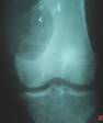
signs were observed. The radiology showed a
metaphysial-diaphysia gap of 7cm in the main line direction, it
had well subscribed internal limits with a bowled over aspect of
the cortical and without any periost reaction (Figure 1).
The biopsy of the tumoural mass recognizes a benign
chondroblastoma. The patient was operated using external access
in front of the tumor, she benefited of curettage of the
tumoural mass with osseous transplant; this last one was
obtained from the iliac bone. The evaluation was favorable with
absence of recurrence in one year recession.
Discussion :
Chondroblastoma is a rare benign
bone tumor; it represents about 1 % of all osseous tumors (1,
2). It is also adolescence tumor, generally occurring between
the ten and the twenty years old and affects mostly male in 60%
of reported cases (3).
The favorite site of this tumor is the superior humeral
epiphysis, superior and inferior femoral, superior humeral of
the tibia (1, 3). The metaphyseal and metaphyseal diaphysia
localization are very rare and exceptional; it only occurs in 2
to 5% of reported series of chondroblastoma (4, 5). Definitely,
about twenty cases were reported since the earliest description
in the literature. The Radiography shows chondroblastoma as
geographic gap, round or oval, regular and well-delimited, it
could squall the bone but it doesnt destroy the cortical. A
cortico-periost reaction could be noticed in half of cases (3).
The positive diagnosis is established using immunohistochemistry
and histological studies (6). The treatment of these tumors is
surgical and consists of curettage of the tumor followed by an
osseous grafting. Reports indicated also the curettage
association with thermic or chemical cauterization using a the
phenol or salty serum (7).
The evolution of the chondroblastoma is mainly dominated by risk
of local recurrence which varies between 5 and 38 % (7). This
recurrence is often accessible for a new curettage-transplant
treatment approach. However, sometimes it is associated to a
tumoural extension in the joint or to adjacent morrow; which
require a less conservative and more extensive surgery, this
would also weight on the functional prognosis.
Reference :
-
Corsat JP, Tomeno B, Forest M,
Vinh TH : chondroblastomes bénins: une revue de 30 cas;
Revue de chirurgie orthopédique, 1989, 75, 179-187
-
Clipatrick SE, Parisen M, Bridge
JA (2002) chondroblastoma in: Fletcher CDM, Unni KK, Mertens
F : world health organisation classification of tumors;
Pathology and genetics of tumors of soft tissue and bonne:
IARC Press, Lyon, pp 241-2
-
Ch. bonard, B de courtivron:
chondroblastome bénin, in les tumeurs osseuses béningnes
de lenfant 1996, 65,69.
-
Dwaik M, Devlin PB.: case report:
metadiaphyseal chondroblastoma; clin. Radiol. 1992 feb; 42
(2): 131-3
-
Pierre Maroteaux, Martine Le
merrer: tumeurs osseuses et processus pseudo- tumoraux bénin,
in M aladies osseuses de l'enfant, 4° edition 2002, 572-610
-
RE, Kurt AM, Sim FH, Unni KK,
Mcleod RA, chondroblastoma, Hum pathol, 1993, 24, 944 949F.
-
accadbled , A brouchet, F.
salmeron, P. davodes, J. P. cahuzac, J. salesde gauzy.
Récidive de chondrobastome agressif, à propos de 2 cas et
revue de la littérature, revue de chirurgie orthopédique 2001;
87, 718-723
|
|
This is a peer reviewed paper Please cite as
: A. Afifi: Exceptional Metaphyseal Diaphysia Localization
of Chondroblastoma
J.Orthopaedics
2006;3(4)e14
URL:
http://www.jortho.org/2006/3/4/e14 |
|
|
ANNOUNCEMENTS |
 |
|

|
|
|
|
|
Arthrocon
2007
CME & Hands on
Workshop
on
Basic Surgical Techniques
in
Arthroplasty
(6th Annual Conference of
COAA)
Demonstrations, Interactive
sessions & Workshop
March 04, 2007
At Port City of Calicut, Kerala, India
Registration
Form
Dr Kishore,
Dept of Orthopaedics,
Medical College, Calicut, Kerala, India
Ph:+91 9895725768
E-Mail:
calicutortho@yahoo.com
|
|
|
Powered by
VirtualMedOnline |
|
