|
Abstract
Subtalar dislocation is the simultaneous
dislocation of the distal articulations of the talus at both the
talocalcaneal and talonavicular joints. It can occur in any
direction and always produce significant deformity. Most common
is the medial dislocation. Less common presentations are
lateral, anterior and posterior dislocations. These dislocations
are associated with osteochondral fractures. Closed reduction
and immobilsation remains the mainstay of treatment. Proper
radiographs and CT scan confirms the post reduction alignment
stability of subtalar joints and intraarticular fracture
fragments. We report a case of antero-medial subtalar
dislocation with no osteochondral fracture fragments in a
25-year-old man.
J.Orthopaedics 2006;3(3)e3
Introduction:
Subtalar dislocations are found rare in
routine orthopedic practice. Many of these dislocations results
from high-energy injuries such as fall from a height, athletic
injuries or a motor vehicle accident. Inversion or eversion
force is dissipated through the weak talonavicular and
talocalcaneal ligaments, which eventually results in subtalar
dislocation.
The head of talus is found medially and the
rest of the foot is dislocated laterally in lateral subtalar
dislocation. The head of the talus is found laterally and the
rest of the foot medially in medial subtalar dislocation. Medial
dislocation has been referred to as an acquired clubfoot,
while the lateral injury is described as an acquired flatfoot.
We present a case of an adult with antero-medial
subtalar dislocation following a fall.
Case report
A 25-year-old man who sustained a fall from
stairs came to our emergency department with pain, swelling
right foot. The foot was diffusely swollen with minimal
laceration and tenting of the skin over the prominent talar
head, which was, felt dorso laterally. The rest of the foot was
found dislocated medially. Posterior tibial artery and dorsalis
pedis artery pulse was not felt due to massive soft tissue
distortion. Radiograph of the right foot showed anterior and
medial subtalar dislocation Fig (1). Doppler ultrasound showed
normal arterial flow in both posterior tibial and dorsal pedis
arteries.
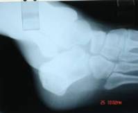
Figure (1) showed the dislocation of
the talo-navicular and subtalar joints. Head of the talus was
seen lying antero-laterally. Normal alignment of calcaneo-cuboid
joint can also be appreciated.
Closed reduction was done under spinal
anesthesia. Firm manual foot traction with counter-traction on
the leg combined with direct digital pressure over the head of
talus aided in smooth reduction, which was associated with an
audible clunk. Post reduction radiographs showed normal and
stable alignment of subtalar and talo-navicular joints with
absence of osteochondral fractures. CT scan confirmed the
absence of osteochondral fractures and the stability of the
subtalar joints. Patient was immobilized in a short-leg
posterior plaster splint for 4 weeks. Vigorous, active exercise
program, progressive weight bearing, active range of motion
exercises to regain subtalar and midtarsal joint motion followed
immobilization. One year after the injury, the patient had a
stable, relatively good functional foot, with minimal pain on
walking on uneven ground.
Discussion :
Subtalar dislocation by
definition has a normal tibiotalar joint. Most dislocations
occur in males (6:1) of early age. Subtalar dislocation can
occur in any direction and always produces significant
deformity. Most commonly (80% to 85%), the foot is displaced
medially with the calcaneus lying medially, the head of the
talus prominent dorsolaterally, and the navicluar medial and
sometimes dorsal to the talar head and neck1,2,3. Less commonly
(15% to 20%), lateral dislocation occurs.
Inversion of the foot
results in a medial subtalar dislocation, while eversion
produces a lateral dislocation. The strong calcaneonavicular
ligament resists disruption, and the inversion or eversion force
is dissipated through the weaker talonavicular and talocalcaneal
ligaments, disrupting these two joints and allowing displacement
of the calcaneus, navicular and all distal bones of the foot as
a unit, either medially or laterally2,3.
The sustentaculum tali acts
as a fulcrum about which the foot rotates to lever apart the
talus and calcaneus in medial subtalar dislocation. The foot
pivots about the anterior process of the calcaneus, again
causing the talus and calcaneus to separate in lateral subtalar
dislocation1,2,3,4.
Rare cases of anterior5 and
posterior1 displacement of the foot after subtalar dislocation
have also been reported. It is important to distinguish the
medial or lateral subtalar dislocations because the method of
reduction is different and the long-term prognosis appears to be
worse with the lateral dislocation.
Between 10% and 40% of
subtalar dislocations are open6. Open injuries tend to occur
more commonly with the lateral subtalar dislocation pattern and
probably as the result of a more violent injury6. Long terms
follow demonstrated very poor results following open subtalar
dislocations.
The keystone of treatment
for all subtalar dislocations is prompt and gentle reduction
under general or spinal anesthesia7. All open injuries must be
thoroughly debrided at the time of reduction, and the wound
should be left open, with delayed primary closure anticipated in
3 to 5 days. Because of the high incidence of associated
articular fracture and associate poor prognosis, CT scan of the
foot and ankle should be obtained after reduction and splinting.
Simple dislocation that are
reduced readily by closed reduction and do not have associated
fracture do very well.1 In approximately 10% of medial subtalar
dislocations and 15% to 20% of lateral dislocations, closed
reduction cannot be achieved3,8. Soft tissue interposition and
bony blocks have been identified as factors preventing closed
reduction. Another common obstruction to closed reduction in
medial dislocations is an impaction fracture of the articular
surface of talus and navicluar7. The most common obstruction to
closed reduction in lateral subtalar dislocation is the
interposed tibialis posterior tendon 8.
Open reduction is done for
irreducible medial, lateral subtalar dislocations and
osteochondral fracture fragments which blocks closed reduction.
Any small, loose articular fracture fragments should be removed,
while large intra-articular fractures should be reduced and
fixed with Kirschner wires or small screws to restore joint
stability and congruity9.
The only consistent
complication in simple uncomplicated dislocations is limitation
of subtalar joint motion, with the occasional associated
symptoms of difficulty in walking on uneven ground and pain in
the foot with weather change2,7. Lancaster and his co-workers
noted a poorer prognosis when there were associated injuries
such as soft tissue injury, open contaminated injuries, extra-articular
fracture, intra-articular fracture, infections, lateral subtalar
dislocations, neglected subtalar dislocations and
osteonecrosis10.
Our patient who had
sustained a fall from stairs came with diffusely swollen foot
with the head of talus felt dorso-laterally and the rest of the
foot dislocated medially as a unit. Radiographs confirmed the
antero-medial subtalar dislocation. There was no associated
osteochondral fracture. Simple closed reduction was successful.
Our literature review showed no reports of isolated antero-medial
subtalar dislocation.
We emphasize the importance of proper
diagnosis and timely management of dislocations around subtalar
joint, as these always produces significant deformity and joint
stiffness. Antero-medial subtalar dislocation is one such type
which is no where mentioned in literature should be carefully
treated and always a high index of suspicion should be kept
about associated osteochondral fractures. CT scan should be done
after reduction to look for the intra-articular fractures of the
subtalar joint. Open reduction is done for irreducible
dislocations and fixations done in large displaced articular
fragments producing subtalar joint incongruity.
Reference :
- DeLee JD, Curtis R. Subtalar dislocation of the foot. J
Bone Joint Surg 1982; 64A:433-437.
- Grantham SA. Medial Subtalar dislocation: five cases with
a common etiology. J Trauma 1964; 4:845-849.
- Heppenstall RB, Farahvar H, Balderston R, et al.
Evaluation and management of subtalar dislocations. J Trauma
1980; 20: 494-497.
- Monson St, Ryan JR. Subtalar dislocation. J Bone Joint
Surg 1981; 63A:1156-1158.
- Inokuchi S, Hashimoto T, Usami N. Anterior subtalar
dislocation: case report. J Orthop Trauma 1997; 11(3):
235-237.
- Golner JL, poletti SC, Gates HS III et al. Severe open
subtalar dislocations: long-term results. J Bone Joint Surg
1995; 77A (7):1075-1079.
- Bohay DR, Manoli A II. Subtalar dislocations. Foot Ankle
Int 1995;16(12): 803-808.
- Leitner B. Obstacles to reduction I subtalar dislocations.
J Bone Joint Surg 1954; 36A:299-306.
- Naranja RA, Monaghan BA, Okereke E et al. Open medial
subtalar dislocation associated with posterior process
fracture of the talus. J Orthop Trauma 1996; 10(2): 142-144.
- Lancaster S, Horowitz M, Alonso J. Subtalar dislocations:
a prognosticating classification. Orthopedics 1985; 8:
1234-1240.
|

