|
*Teppei Suzuki, MD; *Masayoshi Yagi,
MD; *Yasunobu Iwasaki, MD; *Kenji Fujita, MD; *Hideto Maruno, MD;
#Hiroyuki
Fujioka, MD2
*Department of
Orthopaedic Surgery, Shin-suma, Hospital,
Kobe, Japan
#Department of
Orthopaedic Surgery, Kobe University of medicine,
Kobe, Japan
Address for Correspondence
Teppei Suzuki, MD; Department of Orthopaedic
Surgery, Shin-Suma
Hospital,
4-1-6
Isonare-cho Suma-ku
Kobe, 654-0047,
Japan.
Phone: 81 (78) 735-0001
Fax: 81(78) 735-0721
E-mail: teppeisuzuki@hotmail.com
|
|
J.Orthopaedics 2005;2(1)e4
Introduction
Stress fractures are common
entities in intensively trained athletes and are considered to
be a result of repeated application of low-grade stress to the
bones. Stress fractures are most common in the weight-bearing
bones of the lower extremities and the spine, but are rarely
found in the non-weight-bearing bones of the body3,5. In the
following case report, we have reported stress fracture of the
ulnar diaphysis due to
axial
loading during excessive training.
Case report
A 17-year-old volleyball
player had an eight month history of increasing pain of the
ulnar diaphysis. She had started on excessive daily training
program eight months before. Her training required her to stand
on her hands, with a partner holding her up by her ankles (Fig.
1).
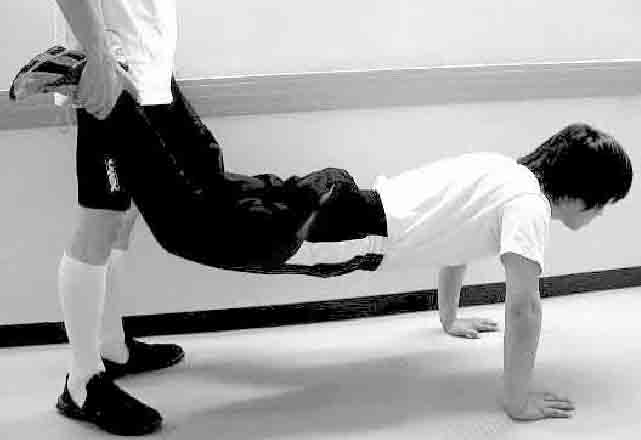
Almost every morning for
four months before playing volleyball, she had to walk on her
hands and sometimes bounce up. The pain in her left forearm
gradually became worse, and she experienced difficulties playing
volleyball. Finally after being hit by a volleyball on the ulna,
she could not continue to play. Almost all activity with the arm
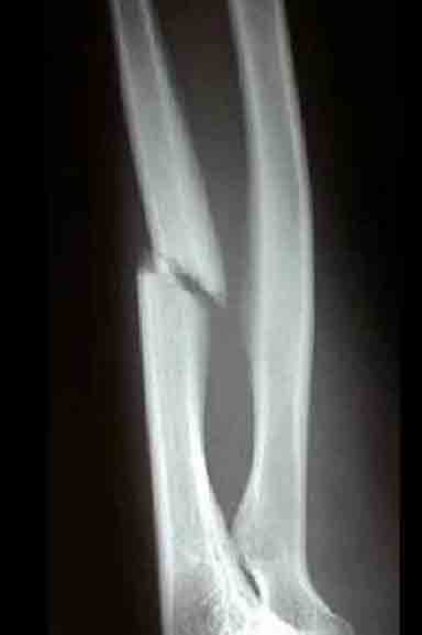
was painful. She was initially diagnosed as having a fresh
fracture of the ulnar diaphysis because radiographs revealed a
clear fracture line in the midshaft of the ulna in July 2002
(Fig. 2).
As a result she was treated
with long arm casting for four weeks. However, after four months
of conservative treatment, the pain continued and gradually
became worse. She was referred to our clinic four months after
the initial diagnosis. On examination, palpation along the left
forearm elicited pain, tenderness, and minimal swelling at the
middle third of the ulna, however there was minimal pain on
pronation and supination of the forearm and flexion and
extension of the wrist and the elbow. There was no malalignment
in the forearm, no wrist joint effusion, no increased warmth,
normal joint motion, and normal health status with no history of
systemic administration of steroids or rheumatologic disorders.
We reviewed the history of the condition again and discovered
that initial pain had actually started four months before the
volleyball struck her, when she had started on excessive daily
training program. The pain in her left forearm gradually became
worse, but she had nevertheless continued this training every
day for four months. Additionally, we discovered that when she
was struck by the volleyball that exacerbated the pain, it was
not with exceptional force but in a manner that might be
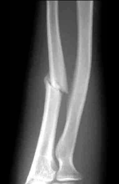
considered normal during play. Thus review of this history and
the plain radiographs showing callus formation of the lower ends
of the ulna in November 2002 (Fig. 3) led us to a diagnosis of
delayed union of a stress fracture of the ulna diaphysis.
We treated the patient with
noninvasive low-intensity ultrasound using SAFHS (Sonic
Accelerated Fracture Healing System; Exogen, Inc., Piscataway,
NJ) with a 100-volt alternating current and ultrasound signal
consisting of a 200-µs burst sine wave of 1.5 MHz repeating at
1.0 kHz. The spatial average, temporal average intensity was
30mW/cm2. The fracture site was exposed to ultrasound for 20
minutes per day. Eight weeks after the start of ultrasound
therapy the patient was pain free.
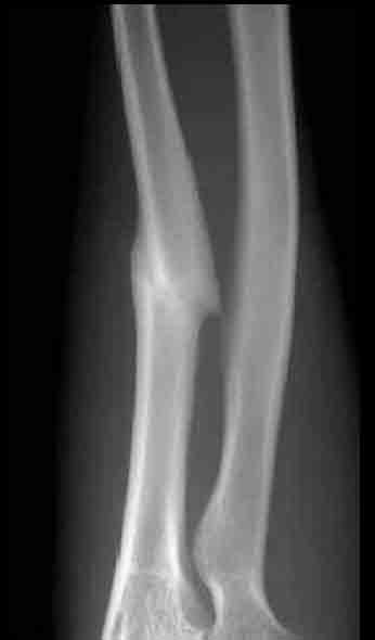
Radiographs showed almost
complete resolution of the fracture and clinical healing
occurred over an eight week period in January 2003 (Fig. 4). The
patient progressed to full workout activities including
competitive volleyball games twelve weeks later.
Discussion
Stress fractures of the
ulnar diaphysis have been described in baseball10 and soft ball
pitchers18, tennis players1,16, body builder11, bowlers7 and
golfer15. The etiology of these fractures has been described as
either a traction injury of the hand flexors and extensors11, or
related to torsional forces associated with excessive pronation
and supination1,18. In this case, the patient had to walk on her
hands leading her wrist to be extended and her forearm pronated
and sometimes bounce up with the wrist ulna deviated, and this
excessive training is very common among all the sports players
in Japanese field training. This type of training required the
patients forearm to be loaded not with torsional, or tractional
force, but with excessive weight-bearing force. This mechanism
of axial loading has previously been reported in a case of
cyclic weight bearing associated with crutch use9. During axial
loading, the radius carries most of the load (82%) and the ulna
carries a smaller load (18%), but the load along the ulna
increases when the wrist is a position of flexion, ulna
deviation, and forearm pronation4. Therefore we hypothesize that
the cause of the fracture was due to axial loading during
excessive muscle training. Low-intensity ultrasound has been
reported to be useful in promoting fracture healing in both
clinical and basic studies8,14,17. In this case, the stress
fracture of the ulna diaphysis had been conservatively treated
for four months, with a final diagnosis of delayed union.
Therefore standard procedures such as resting of the affected
limb were non-effective in this case. The athletes rapid return
to full sporting activity may be attributed to the acceleration
of healing due to low-intensity ultrasound therapy13. It has
been estimated that between five and ten percent of all
sports-related injuries involve stress fractures11 and a good
proportion of these result in delay or non-union. For example,
Hulkko and Orava estimated delay or non-union in ten percent of
stress fractures in Finland12. The reason for these poor result
are two fold: delayed diagnosis due to late consultation of
expert physicians, and/or too short a rest from hard physical
activity. Furthermore in many cases, the diagnosis is difficult
and repeated clinical, radiological examinations are necessary.
Emphasis should therefore be placed on the earliest possible
diagnosis and provision of effective primary treatment. The
mechanism of stress in present case is only speculative, but it
suggests the possibility of stress fractures of the ulna
diaphysis in individuals participating in excessive training
regimens such as that outlined in this paper.
REFERENCES
1. Bollen SR Robinson DG
Crichton KJ et al: Stress fractures of the ulna in tennis
players using a double-handed backhand stroke Am J Sports Med
21: 751-752, 1993
2. Brand JC Jr Brindle T Nyland J et al: Does pulsed low
intensity ultrasound allow early return to normal activities
when treating stress fractures? A review of one tarsal navicular
and eight tibial stress fractures Iowa Orthop J 19: 26-30, 1999
3. Brooks AA: Stress fractures of the upper extremity. Clin
Sports Med Jul 20(3):613-620, 2001
4. Clark R J Sizer PS Jr Slavterbeck: Stress fracture of the
ulna in a male competitive polo player Am J Sports Med 30(1):
130-132, 2002
5. Courtenay BG, Bowers DM: Stress fractures: clinical features
and investigation. Med J Aust Aug 6;153(3): 155-156, 1990
6. Ekenstam FW Palmar AK Glisson RR: The load on the radius
and ulna in different positions of the wrist and forearm A
cadaver study Acta Orthop Scand 55:363-365, 1984
7. Escher SA: Ulnar diaphyseal stress fracture in a bowler Am J
Sports Med 25(3):412-413 1997
8. Fujioka H, Tsunoda M, Noda M, et al.: Treatment of ununited
fracture of the hookof
the hamate by low-intensity pulsed ultrasound. A case report. J
Hang Surg Am
|

