Address for communication
Dr Sreesobh K V
Department of Orthopaedics
Amrita institute of medical science and research centre.
AIMS Ponekkara P.O, Kochi, Kerala, India. 682041
Email: drsreesobh@gmail.com,
jaithilak@amrita.aims.edu, kshvshn381@gmail.com, nphilip1@gmail.com
Abstract:
Introduction: femoral component rotation in total knee arthroplasty has crucial role in ligament balancing, patellofemoral mechanics and long term survival. Several axes are described for aligning the femoral component in appropriate rotation mainly posterior condylar axis, transepicondylar axis and anteroposterior axis (Whitesides). Computer assisted surgical navigation systems has been developed in an attempt to help the surgeons to align the components more accurately. The purpose of this study is to find out if the 3 degree external rotation to the posterior condylar axis by mechanical guides matches with the computer navigation values and also to find out the correlation between these three axes values projected by navigation
Materials and methods: This study included 168 primary total knee replacements in 132 patients from January 2008 to June 2010 in Indian patients performed with computer navigation. The study has excluded non osteoarthritic and grossly deformed knees.
During total knee arthroplasty, the anteroposterior cutting jig was placed at a 3 degree external rotation to the posterior condyles by mechanical guides. The navigation tracker was placed on this manually placed jig, and its relation to the TEA, PCA and Whiteside’s line provided by the BrainLab software were noted. Comparison and statistical evaluation of these values were done.
Results: The navigated PCA values were corresponding to the fixed 3 degree external rotation guide. The TEA and Whiteside’s line showed significant deviation from the expected value (zero) when the component was positioned with reference to posterior condyles by jig. The study demonstrates no significant statistical correlation between PCA, TEA and Whiteside’s line.
Conclusion: The study concludes no close correlation between three commonly used referencing systems for the rotational alignment of the femoral component by navigation in our set of patients. More cadaver and CT studies are required to determine the relationship between these axes in Indian population and to find out which axis would provide the most appropriate femoral component rotation.
Key words: total knee arthroplasty, femoral component rotation, computer navigation, axis
Introduction
Total knee arthroplasty has demonstrated long term success in the treatment of advanced osteoarthritis and other disabling conditions of the knee joint. One of the important parameters for achieving this success is in perfect alignment of tibial, femoral and patellar components. Obtaining proper femoral rotational alignment of the femoral component during total knee arthroplasty is critical and challenging step that influences tibiofemoral kinematics 1, patellofemoral tracking and soft tissue balancing especially coronal laxity in flexion. Excessive external rotation of femoral component is associated with undue varus laxity in flexion (medial condylar lift off) while excessive internal rotation leads to lateral condylar lift off and also associated with patellar instability and patellofemoral complications 2 3 4.
Several reference axes have been proposed to establish correct rotational alignment of the femoral component that includes posterior condylar axis (PCA)5, 6, transepicondylar axis (TEA) 6,10, 11 and the anteroposterior axis (Whitesides line).7, 8 The TEA is approximately 3 degrees of external rotation in relation to the PCA6 and Whitesides line is perpendicular to TEA7, 8. The PCA may be misleading because of the degeneration of the posterior femoral condyles especially in valgus knees.9, 10, 11. Aligning component parallel to TEA has been shown to approximate the flexion – extension axis of the knee better17, optimize patellofemoral tracking and increase in the longevity of the implants 10. Debate continues about how accurately the TEA can be located since landmarks on the distal femur are highly variable6, 12, 13. The Whiteside’s axis is easily identified during surgery7, 8. But, there exists great interobserver variability and it may also lead to malrotation due to erosion of anterior part of femoral condyles14. In addition, there are reports stating differences in the morphology of distal femur in various populations14, 15, 16.
Computer assisted surgical navigation systems has been developed in an attempt to help the surgeons to align the components more accurately than what is possible by traditional mechanical guides18 19, 20. There are several reports that demonstrate better component positioning in frontal and sagittal planes but the controversy exists regarding whether computer navigation improves femoral rotation 20, 21, 22, 23.
The purpose of this study is to find out if the 3 degree external rotation to the posterior condylar axis by mechanical guides matches with the computer navigation values and also to find out the correlation between these three axes values projected by navigation.
Materials and methods:
This study included 168 primary total knee replacements in 132 patients (36 received bilateral knee replacements) from January 2008 to June 2010 for primary osteoarthritis. 31 were males and 101 were females. All knees were operated by a single surgeon (the senior author). These patients received LPS (Zimmer, Warsaw, Indiana) and PFC CS & CR (Depuy, Warsaw, Indiana) performed with computer navigation (BrainLAB, Feldkirchen, Germany). The study has excluded patients of non-Indian origin, those with rheumatoid arthritis, valgus deformity of more than 10 degrees and varus more than 25 degrees.
Intraoperatively, the posterior condyles, epicondyles and Whitesides line are registered along with other reference points to the BrainLAB computer. The TEA was assessed by identifying the lateral epicondyle and most prominent part of the medial epicondyle. The Whitesides line was marked from the apex of the intercondylar notch to the deepest point of the trochlear groove. The anteroposterior cutting jig was placed at a 3 degree external rotation to the posterior condyles. The navigation tracker was placed on this manually placed jig, and its relation to the TEA, PCA and Whitesides line provided by the BrainLAB software were noted. Comparison and statistical evaluation of these values were done.
Results:
The navigated PCA values were corresponding to the fixed 3 degree external rotation guide (Z score = 1.31). 89 knees (52%) came within a reference range of 2 to 4 degree of external rotation. 11 (6.5%) knees were deviated more than 3 degrees from the expected (3 deg. External rotation) (Figure 1).
The TEA and Whiteside’s line showed significant deviation from the expected value (zero) when the component was positioned with reference to posterior condyles by jig. (Z score for TEA is 5.04 and Whiteside’s line is 11.16 with a p value of less than 0.001) (Figure 2 & 3). When analyzing TEA, only 30 (17%) femoral components were aligned within 1 degree of internal or external rotation. There were 70 knees (41 %) in which the femoral component was deviated by more than 3 degrees.
Whitesides line showed the maximum deviation to the jig based PCA. The majority of the components turned out to be in internal rotation with respect to Whitesides line. 98 knees (58.3 %) were deviated by more than 3 degrees.
The Table 1 summarizes the results -
Table 1:
|
Referral value |
+/- 1 |
+/- 2 |
+/- 3 |
> +/- 3 |
PCA |
3 ER |
89 |
133 |
156 |
11 |
TEA |
0 |
30 |
60 |
98 |
70 |
Whitesides |
0 |
19 |
47 |
70 |
98 |
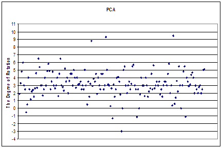
Figure 1: The femoral rotational alignment with the respect to the PCA (navigational) when the component is placed at the 3 degrees of external rotation manually by jig.
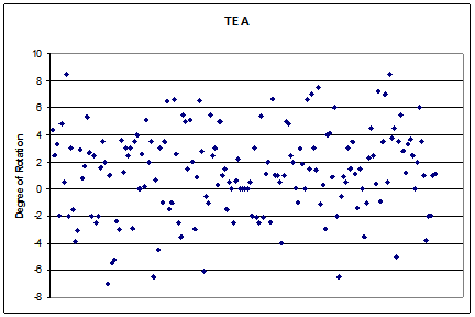
Figure 2: The femoral rotational alignment with the respect to the TEA when the component is placed at the 3 degrees of external rotation manually by jig.
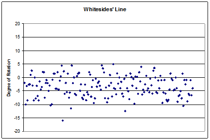
Figure 3: The femoral rotational alignment with the respect to the Whitesides’ Line when the component is placed at the 3 degrees of external rotation manually by jig.
Pearson correlation:
TEA - PCA - 0.105
TEA - Whitesides - 0.189
PCA - Whitesides - 0.074
The study demonstrates no significant statistical correlation between PCA, TEA and whitesides line.
Discussion
Various alignment axes are utilized to implant the femoral component in proper rotation. Most systems using posterior condylar reference recommend three degree external rotation for aligning femoral component. It is not clearly known whether this practice can reproduce the appropriate femoral rotation.
In the present study the navigated posterior condylar axis matched to the fixed 3 degree jig. But the critical question is whether we are achieving proper rotational alignment with respect to TEA and Whitesides by referencing the posterior condylar axis. This is important as some authors recommend TEA as the most ideal reference axis whilst others recommend Whitesides line. However, a study on cadavers reports all techniques resulted in highly variable rotational alignment23. A study by W J Hozack et al (mostly on Caucasian race) has shown close correlation between the PCA, TEA and Whitesides line24. In our study, no such correlation was found. If TEA is assumed as the best axis, then 41 % of the knees would be malrotated (>3 degrees) by implanting the prosthesis with jig. This number increases to 58 % for the Whitesides. Conversely, if PCA is taken as the more accurate axis, then referencing the TEA and Whitesides would result in femoral component malrotation.
The limitations of the study: All the cases in this series were operated by single surgeon, so the inter- observer variations in registering anatomical landmarks were not obtained. It is not a cadaver study, which would render more accurate landmark identification. Moreover; no post operative scannograms were done to find out the exact rotation of the femoral component.
Conclusion: -
The study concludes no close correlation between three commonly used referencing systems for the rotational alignment of the femoral component by navigation in our set of patients. The navigated values of PCA could be matched to the jigs referencing the posterior condyles. More cadaver and CT studies are required to determine the relationship between these axes in Indian population and to find out which axis would provide the most appropriate femoral component rotation.
Competing interest:
The authors did not receive any outside funding or grants in support of their research or preparation of this work. Neither they nor a member of their immediate families received payments or other benefits or a financial or non-financial commitment or agreement to provide such benefits from a commercial entity.
Authors’ contribution:
Dr Sreesobh K V was substantially involved in the design of the study, acquisition of data, analysis and interpretation of data and in drafting of the manuscript. Dr Jai Thilak contributed to the conception and basic framework of the study, critically analyzed and evaluated the project at each stage and critically revised the manuscript. Dr Keshav Shenoy participated in the acquisition and analysis of the data. Dr Nileena Philip helped in the data analysis and drafting of the manuscript. All authors have read and approved the final manuscript
Acknoledgment:
We acknowledge the statistic help and guidance from Prof Sundaram, Amrita Institute of Medical Sciences.
References
1.Miller MC, Berger RA, petrella AJ, Karmas A, Rubash HE,. Optimising femoral component rotation in total knee arthroplasty. Clin Orthop Relat res. 2001; 392:38-45
2.Akagi M, Matsusue Y, Mata T, Asada Y, Horiguchi M, Iida H, Nakamura T. Effect of rotational alignment on patellar tracking in total knee arthroplasty. Clin Orthop Relat Res 1999;366:155–63
3. Rhoads DD, Noble PC, Reuben JD, Mahoney OM, Tullos HS. The effect of femoral component position on patellar tracking after total knee arthroplasty. Clin Orthop Relat Res 1990; 260:43–51.
4. Singerman R, Pagan HD, Peyser AB, Goldberg VM. Effect of femoral component rotation and patellar design on patellar forces. Clin Orthop Relat Res 1997; 334:345–53
5. Matsuda S, Matsuda H, Miyagi T, Sasaki K, Iwamoto Y, Miura H. Femoral condyle geometry in the normal and varus knee. Clin Orthop Relat Res 1998;349:183–8.
6. Berger RA, Rubash HE, Seel MJ, Thompson WH, Crossett LS. Determining the rotational alignment of the femoral component in total knee arthroplasty using the epicondylar axis. Clin Orthop Relat Res 1993; 286:40–7.
7. Arima J, Whiteside LA, McCarthy DS, White SE. Femoral rotational alignment, based on the anteroposterior axis, in total knee arthroplasty in a valgus knee. A technical note. J Bone Joint Surg Am 1995;77:1331–4.
8. Whiteside LA, Arima J. The anteroposterior axis for femoral rotational alignment in valgus total knee arthroplasty. Clin Orthop Relat Res 1995;321:168–72.
9. Griffin FM, Insall JN, Scuderi GR. The posterior condylar angle in osteoarthritic knees. J Arthroplasty 1998;13:812–5.
10. Olcott CW, Scott RD. A comparison of 4 intraoperative methods to determine femoral component rotation during total knee arthroplasty. J Arthroplasty 2000;15:22–6.
11. Yoshino N, Takai S, Ohtsuki Y, Hirasawa Y. Computed tomography measurement of the surgical and clinical transepicondylar axis of the distal femur in osteoarthritic knees. J Arthroplasty 2001;16:493–7.
12. Katz MA, Beck TD, Silber JS, Seldes RM, Lotke PA. Determining femoral rotational alignment in total knee arthroplasty: reliability of techniques. J Arthroplasty 2001;16:301–5.
13. Kinzel V, Ledger M, Shakespeare D. Can the epicondylar axis be defined accurately in total knee arthroplasty. Knee 2005;12:293–6.
14. Nagamine R, Miura H, Inoue Y, Urabe K, Matsuda S, Okamoto Y, et al. Reliability of the anteroposterior axis and the posterior condylar axis for determining rotational alignment of the femoral component in total knee arthroplasty. J Orthop Sci 1998;3:194–8.
15. Yip DK, Zhu YH, Chiu KY, Ng TP. Distal rotational alignment of the Chinese femur and its relevance in total knee arthroplasty. J Arthroplasty 2004;19:613–9.
16. Mullaji AB, Sharma AK, Marawar SV, Kohli AF, Singh DP. Distal femoral rotational axes in Indian knees. J Orthop Surg. 2009; 17: 166-9.
17. Churchill DL, Incavo SJ, Johnson CC, Beynnon BD. The transepicondylar axis approximates the optimal flexion axis of the knee. Clin Orthop Relat Res 1998 ; 356: 111-8
18. Stulberg SD, Loan P, Sarin V. computer assisted navigation in total knee replacement.: results of an initial experience in thrity five patients. J Bone Joint Surg Am. 2002; 84 Suppl 2 : 90-8
19. David Stulberg S. How accurate is current TKR instrumentation. Clin Orthop Relat Res. 2003; 416: 177-84
20. Chauhan SK, Scott RG, Breidahl W, Beaver RJ. Computer assisted knee arthroplasty versus a conventional jig based technique. A randomized, prospective trial. J Bone Joint Surg Br . 2004; 86: 372-7
21. Stockl B, Nogler M, Rosiek R, Fischer M, Krismer M, Kessler O. Navigation improves accuracy of rotational alignment in total knee arthroplasty. Clin Orthop Relat Res. 2004; 426: 180-6
22. Siston RA, patel JJ, Goodman SB, Delp SL, Giori NJ. The variability of femoral rotational alignemtn in total knee arthroplasty. J Bone joint Surg Am. 2005; 87: 2276-80
23. Matziolis G, krocker D, Weiss U, Tohtz S, Perks C. A prospective, randomized study of computer assisted and conventional total knee arthroplasty. Three dimentional evaluation of implant alignment and rotation. J Bone Joint Surg Am. 2005; 89:236-4
3
24. Restrepo C, Hozack WJ, Orozco F, parvizi J. Accuracy of femoral rotational alignment in total knee arthroplasty using computer assisted navigation. Comp Aided Surg.2008; 13: 167-172
|

