Address for Correspondence
Address for correspondance:
Dr. Nilesh Giripunje. J J HOSPITAL MUMBAI.
email: nilesh9826@yahoo.co.in
Telephone: 09869109826.
Abstract:
Major disadvantage of Transtrochanteric Rotational Osteotomy (TRO) for osteonecrosis of head is high operative complication rate like neck fracture because of on going femoral head necrosis, progressive varus deformity, collapse of head and long postoperative recovery period. We describe here two cases of stress fracture of the femoral head after TRO. 18 patients underwent TRO from January 2000 to August 2007, 17 male and 1 female and age ranges from 20 to 47 years. Out of 16 patients treated by sugioka osteotomy 2 patients who were manual worker and mountain climber sustained femoral head fracture after continuous mountain climbing for 7 to 8 hours, one and half to three years after successful sugioka operation. Both patients had no history of precipitating trauma.
Key words: Osteonecrosis of femoral head, Alcoholic, Transtrochanteric rotational osteotomy, Mountain climbing, Femoral head fracture.
J.Orthopaedics 2011;8(2)e4
INTRODUCTION:
Hip arthritis is a serious complication of osteonecrosis of femoral head. Middle aged (1) group people are more involved. Main aim of osteotomy is to reposition necrotic anterosuperior part of femoral head to a non weight locale. To get a good result femoral head should have at least one third of normal articular surface pre operatively. The subchondral portion is a common site of fracture in osteonecrotic femoral head, however another common site for fracture is the junction between necrotic bone and reparative bone, and this junction is in the sub capital area when osteonecrosis involves the whole femoral head (10).
Femur neck fracture can develop after Sugioka transtrochnateric roatational osteotomy(TRO) with incidence of 1-15% ( sugioka, shon, sugano). To the author’s knowledge, however no case with femoral head fracture after Sugioka transtrochnateric roatational osteotomy has been reported. The authors report a rare case of femoral head fracture after TRO and discuss the importance of the period of postoperative rehabilitation after TRO.
CASE REPORT:
PATIENT 1: 40 years male textile worker by occupation, a mountain climber every weekend and alcoholic since last 15 years came to us with diagnosis of osteonecrosis of right femoral head, At the time of diagnosis patient was in stage 1 of Ficat Arlet classification, and according to Sugano’s classification (8) it was of Type C1, with terminal restriction of movements, and preoperative HHS of 90. Patient got operated 10 days after diagnosis with above method, and osteotomy site was fixed with four 6.5mm cannulated cancellous screws. Patient was kept in skin traction for 4 weeks with intermittent Quadriceps and active Range of Motion (ROM) exercises, non weight bearing crutch walking was allowed after 6 weeks for period of 3 months, followed by partial weight bearing for another 3 months, crutch walking was continued till 4 to 6 months after surgery (3). Post operative HHS was 96. Patient was allowed full weight bearing walking after 9 months and mountain climbing was allowed 10 months after the surgery. Radiograph taken at 8 months post op shows union of osteotomy site. He was called for follow up every 6 week till first 3 months and there after 3 months interval (Fig 1). Almost after 3 years patient came with chief complaints of acute pain in (R) hip joint after doing mountain climbing continuously for 8 to 10 hours, unable to walk, and pain ful range of movement. CT scan and roentgenogram done, showed vertical undisplaced fracture of anterolateral portion of femoral head. Patient was kept in Bucks traction for 1 month and then discharged with non weight bearing crutch walking for 4 months, and then partial weight bearing for another 2 months. Full weight bearing was begun after a total of 6 months. Every 15 days interval, patient was called up for follow up and routine hip roentgenogram was done. CT scan was done at 3 months to evaluate the fracture healing Last follow up roentgenogram done 13 months after detection of head fracture showed healed fracture (Fig 2).
PATIENT 2: 46 years male labor by occupation and a mountain climber everyday for 2 to 3 hours and alcoholic since last 20 years with diagnosis of osteonecrosis of right femoral head. Patient at the time of diagnosis was in stage 2 of Ficat Arlet classification and according to Sugano’s classification (8) it was of Type C2 and pre operative HHS was 85, with terminal restriction of movements. Sugioka osteotomy was done and osteotomy site was fixed with DHS and 125º side plate. Similarly patient was kept non weight bearing for 3 months and partial weight bearing for 3 more months. All routine work and full weight bearing walking was allowed 8 months after the surgery but mountain climbing was delayed for 3 more months (Fig 3). Radiograph taken before allowing patient mountain climbing shows union of osteotomy site. Around one and half year after sugioka osteotomy patient came with history of mountain climbing for continuously 6 to 7 hours with pain in Right hip, not able to walk and painful ROM.CT scan and roentgenogram done shows displaced vertical fracture of anterolateral femoral head. Patient was kept in Bucks traction for 15 days and then discharged with non weight bearing crutch walking, and called for follow up every 2 weeks. After 5 months of conservative management CT scan done showed further displacement of fracture site (Fig 4), so conservative treatment was abandoned and Total Hip Arthroplasty was done subsequently.
PATIENT3: 39 years old male patient came with diagnosis of osteonecrosis of Right femoral head, in a Ficat Arlat stage 2 and Sugano’s Type C2. Patient was alcoholic for 15 years and mountain climber since 15 years. Pre operative HHS was 90 with terminal restrictions of movements. Patient got operated and Sugioka osteotomy was done and osteotomy site was fixed 125° DHS. Patient was kept non weight bearing for 3 months and partial waeght for 3 more months and full weight bearing was started 6 months after the operation. Mountain climbing was allowed 8 months after the surgery. 1years after allowing mountain climbing patient came with history of mountain climbing for 5 to 6 hours continuously and not able to walk after that because of pain. Radiograph and CT scan did shows displaced fracture of femoral head. After few days patient THA was done.
DISCUSSION
Complication of femoral head fracture is not yet reported but complications like collapse of necrotic segment, progressive varus deformity, and femoral neck stress fracture are not uncommon (3) Masafumi Homma (2) had following early complications in his study of Sugioka Osteotomy like fracture of lesser trochanter, subtrochanteric fracture and few late complications like deep infection, femur neck fracture, delayed union, nonunion of greater trochanter. Sugioka et al reported femoral neck fracture in four of 474 hips after TRO(4) , ,even Sugano et al (11) reported 6 cases of femoral neck fracture in his case study of 41 hips after TRO, while Shon et al (3)reported 3 cases of neck fracture out of 64 hips. Subchondral insufficiency fracture of femoral head in osteonecrosis is reported by Sudo A (5). For subchondral insufficiency fracture Young-Min Kim (10) listed various causes like osteoporosis, osteogenesis imperfect, pagets disease, hyper parathyrodism, osteomalacia, scurvy, rheumatoid arthritis, post irradiation, fibrous dysplasia and osteopetrosis. Once osteonecrosis involves entire head subcapital femoral head fracture at the junctional area between necrotic and reparative bone is common, similarly subchondral fracture after extensive osteonecrosis are not uncommon (9).
As such CT scan reveals more subchondral fractures in osteonecrosis of the femoral head than unenhanced radiography or MR imaging (6). We used CT scan to know exact fracture anatomy and further to judge the collapse of head. The necrotic portion of femoral head was located inferomedially and the fracture detected was superolaterally. The two were not close to each other. (Fig 1C, Fig 2C). During mountain climbing hip frequently goes in flexion, abduction and external and internal rotation. During climbing up at one point whole of body weight comes on single hip, and it is more or less like stance phase of normal gait were hip has to bear 3 to 6 times the body weight (7). As the fracture in our cases was away from the area of necrosis, this fracture may have been caused by undue increased stresses for 7 to 8 hours continuously in the region of femoral head where osteonecrosis was not apparent on initial MRI. In both the patients fracture line is almost similar in location and direction.
Structural breakage of the femoral head in osteonecrosis is initiated by focal resorption of the subchondral bone plate during the repair phase. The triggering factor for fracture to occur remains unknown. The authors conclude that patients who have undergone sugioka osteotomy should avoid any kind of heavy work such as mountain climbing etc should be avoided for a 4 - 5 years after TRO
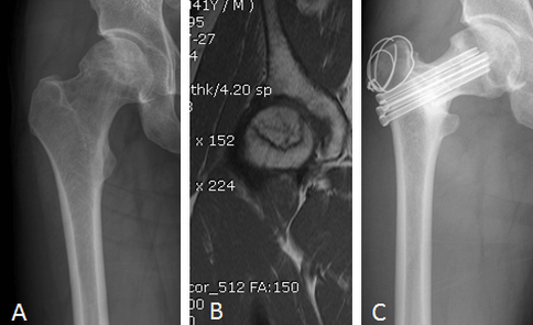
Fig 1:(A) and (B): AP radiography of Right hip and MRI shows Sugano’s Type C2 Osteonecrosis of Right femoral head, Lesion extends lateral to acetabular edge.
(C)situ with complete union and remodeling of osteotomy site, no collapse of femoral
head, with healed osteonecrosis and maintain joint space.
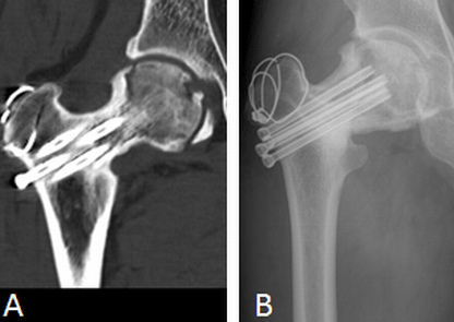
Fig 2:(A): CT scan taken after the complaints shows vertical undisplaced fracture involving
anterolateral portion of femoral head.
(B): AP radiography taken 13 months after fracture shows minimal collapse of head
anterosuperiorly Without collapse of osteotomy site as compared to previous
radiographs with healed fracture and spherical femoral head.
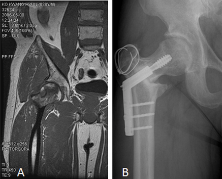
Fig 3: (A): MRI Shows Sugano’s type C2 Osteonecrosis of the Right femoral head involving anterolateral Portion.
(B) A P Radiography of Right hip joint taken 10 months after the Sugioka operation
shows implant insitu, with Complete union and remodeling of osteotomy site, with
no collapse of femoral head, and maintain joint space.
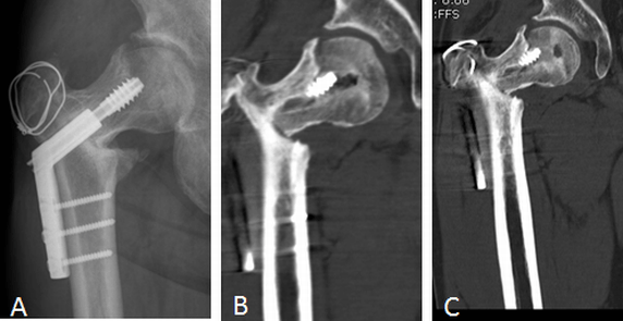
Fig 4: (A): Immediate AP radiography shows fracture of the femoral head.
(B): CT scan taken after the complaints shows vertical minimal displaced fractur
involving anterolateral portion of the femoral head.
(C): CT scan taken 4 months after conservative treatment shows further displacement
of the fracture site as compared to Fig 4(B).
REFERENCES
1. Transtrochanteric Rotational Osteotomy, Section After Sugioka.Vojtassak j, jakubik T. bratisl lek listy 2001; 102 (11): 536-540
2. Sugioka Osteotomy. Clinical case presentation ( October 26,1995 ) Masafumi Homma, Richard Bowen
The Alfred I.Dupont Institute Wilmington,Delaware
3. Transtrochanteric Rotational Osteotomy For Nontraumatic Osteonecrosis Of The Femoral Head In Young Adult. Sandeep Biswal, Sunit Hazra, Ho Hyun Yun, Chang Yong Hur, Won Yon Shon. CORR 10.1007/s
11999-008-0696-3.
4. Transtrochanteric Anterior rotational Osteotomy For Idiopathic And Steroid Induce Necrosis Of The Femoral Head:Indication And Long Term Results . Sugioka Y,Hotokebuchi T, Tsutsui H. CORR 1992 April, (277):111-20
5. Bilateral Subchondral Insuffency Fracture Of Femoral Head. Sudo A, Hasegawa M, Kato K, Uchida A :Orthopaedics 2008 April,31(4):399
6. Subchondral fracture in osteonecrosis of femoral head: comparison of radiographic,CT, and MR imaging .
Kathryn stevens,corline tao,shi-UK lee,Natalie salem. AJR AM J Roentgenol,2003 feb; 180(2):363-8.
7. Campbells Operative Orthopaedic. Ninth Edition Volume One
8. The 2001 revised criteria for diagnosis,classification,and staging of idiopathic osteonecrosis of the femoral head. N.sugano,t.atsumi..j orthop sci(2002) 7:601-605
9. Subcapital Fracture Associoated With Extensive Osteonecrosis Of The Femoral Head.Byung Woo Min,MD, Kyung-Hoi Koo,MD….CORR Number 390,pp 227-231.
10. Pathological fracture of the femoral neck as the first manifestation of the osteonecrosis of the femoral head.young-kim and hee joong kim.j ortho sci (2000) 5:605-609
11. Rotational osteotomy for non-traumatic avascular necrosis of the femoral head N Sugano, K Takaoka, K Ohzono, M Matsui, M Saito, and S Saito. JBJS BRI. VOL 74_B Issue 5, 734-739.
|


