| ORIGINAL
ARTICLE |
|
Short Segment Anterior Correction
Of Thoracic Scoliosis With Single Solid Rigid Rods.( In
Adolescent Idiopathic Scoliosis) |
|
Gopinathan P*,Anwar Marthya,Kumaran C
M,Chang Whan Han, Kishore Puthezeth,Sibin surendran
*
Department of Orthopedics, Medical College,Calicut,Kerala,india
Address for Correspondence:
Dr Gopinathan P
Department of Orthopedics,
Medical College,Calicut,Kerala,india
|
|
Abstract:
Study design:
Retrospective case series review
Objectives:
To evaluate the results of single solid rigid rod anterior
instrumentation in thoracic scoliosis (Adolescent idiopathic
scoliosis).
Summary of
background data: Scoliosis surgery is usually performed
through posterior approach. But anterior correction is more
physiological in terms of amount of correction and the number of
segments saved. Some authors have reported that the anterior
correction is kyphogenic and may lead to implant failure, loss
of correction and pseudarthrosis, but this is extremely rare
with the current system of implants.
Patients and
Methods: 20 patients with adolescent idiopathic scoliosis
with average 75° cobbs angle, and with an average age of 14.5
were operated with a single rigid anterior rod screw systems
were included in this study. All patients were available for
follow up. The average follow up was 3 years. Ranges from 1.5
years to 4.5 years. In 5 patients distal most vertebrae was
excluded from fusion (short segment fusion). Radiographic
evaluation of both pre op and post op were made to evaluate the
sagital and coronal alignment of the spine. SRS outcome
instrument charts were reviewed in all patients.
Results:
The average correction rate of scoliosis was 80% over
uninstrumented level and 90% over instrumented level. The
average pre-op Thoracic kyphosis was 9° corrected to 12° Post
op. There were no implant related complications. Compensatory
curves spontaneously improved by an average of 42% for upper
curves and 60% for lower curves. Fusion could be achieved in
all patients at latest follow up. One compensatory curve
progressed in thoraco-lumbar region and was managed with Knight
Tailors brace and eventually improved. Thoracic, thoracolumbar
and lumbar sagital alignment improved in all patients. Surgery
did not induce kyphosis in any of the patients. SRS outcome
instrument chart review showed that overall satisfaction score
4.2/5, self image score of 4.5/5 with average 80% overall
score. Lumbar lordosis improved to 50° to 30° after surgery.
Trunk shift improved from 30 mm to 6 mm. Improvement of the end
vertebra tilt was by 94% in the instrumented group of curves and
80% in the uninstrumented group of curves. The apical vertebra
rotation was corrected by 86%. Pre op kyphosis in the thoracic
region was 7° and post op 12° . Rib hump angle was corrected by
60%.
Conclusion:
The results of anterior single rigid rod instrumentation gives
excellent results with 100% fusion rate. It gives perfect
saggital and coronal plane alignment with good rotary
correction. Physiologic lumbar lordosis could be achieved with
normal lumbar Lordosis . There is no evidence of excessive
kyphosis, implant failure, pseudarthrosis or gross loss of
correction at latest follow up.
J.Orthopaedics 2008;5(2)e11
Introduction:
Surgical
correction of adolescent idiopathic scoliosis continues to
improve with the emergence of better implants. The traditional
method of correction of scoliosis is by posterior approach, but
anterior correction is more physiological in terms of curve
correction, fusion and maintenance of correction. There are
only scanty reports in English literature regarding anterior
correction of thoracic scoliosis; even though anterior
correction of thoracic and thoraco-lumbar scoliosis is a well
established procedure. Anterior fusion with special
instrumentation was first introduced by Dwyer et al in 1969
(1). It merely produced compressive force between vertebral
bodies, but pseudarthrosis rate was high(1). In thoracic
scoliosis, posterior instrumentation has been the main component
of surgical management. Harrington (2) introduced spinal
instrumentation in late 1950s. The principle of distraction
often reduced the normal thoracic kyphosis (3). Further
improvements were made by Cotrel and Doubassette instrumentation
(4,5). Zeilky instrumentation (6,7,8,9) has been associated
with high rate of implant failure and pseudarthrosis. Kaneda
anterior instrumentation was developed with excellent results
(10,11). The advantages of anterior correction in scoliosis
involves better deformity correction, correction of deformity at
the site of deformity and maximum motion segment preservation
by short segment fusion. The other anterior instrumentation
system used in scoliosis include, Texas Scotish Rite system and
ISOLA system (10,12,13). Even with rigid systems like this,
there are reports of implant failure and pseudarthrosis (14).
In this study, vertebral screws with single solid rigid rod
anterior instrumentation is used for anterior thoracic scoliosis
correction.
Aim :
The current
study was done to asses the efficacy of single rigid rod,
anterior correction of thoracic scoliosis in terms of deformity
correction, fusion and loss of correction on follow up.
Patients and Methods :
20 consecutive
patients with adolescent idiopathic scoliosis irrespective of
sex were operated in the Spine surgery division of Dept of
Orthopaedics, in Calicut Medical College south India during the
year 2001 to 2006 were reviewed. Only thoracic curves between
the level of T4 to T11 were selected for the study. The
inclusion criteria were adolescent idiopathic scoliosis less
than 19 years of age, single rigid rod anterior spinal
instrumentation with fusion, thoracic curves, and minimum follow
up of one year. Average age was 14.5. All patients were
treated with stand alone single rigid anterior rod fixation
without tetra-spikes. Thoracic curves are selected with at
least 60% flexibility of secondary curves were selected. All
patients had thoracic major curves. King classification was
followed in this study (15).
Radiographic
evaluation was done pre operative post operative and follow up.
Standing AP, Lat stress views,stretch views and rib hump views
were taken in all patients. The radiographic measurements
involved frontal (Major Thoracic, Major T-L, Compensatory TL and
lumbar) Sagital cobbs angle T2 T12 for thoracic kyphosis,L1
L5 for lumbar lordosis,
T-L junction
alignment was measured fromT10-L3.Coronal and sagital trunk
translation was measured radio- graphically by measuring the
distance from the central sacral line and from the middle of the
C7 body respectively
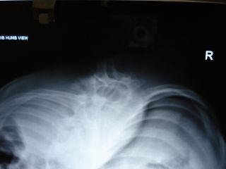
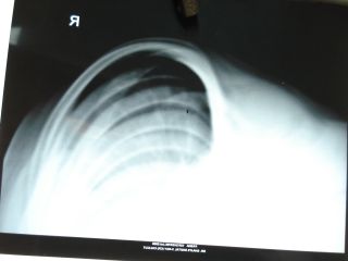
Stagnara views
were taken in all patients to asses the rotational element. Rib
hump views were taken in all patients to assess the torso
rotation. All x-rays were taken preoperatively and post
operatively.
Selection of
fusion levels was done in neutral standing and bolster view in
lying down position,(it is taken in the lateral decubitus
position with a bolster under the convex side and AP view of the
spine is taken). Fusion was done from end vertebra to end
vertebra. The distal end vertebra was saved in some patients
based on horizontalization of distal end vertebra on a stretch
film, which normally would have been instrumented in a posterior
approach. In major thoracic curves the TL or L curves were taken
as compensatory curves. In major TL curve Thoracic curve is
taken as compensatory which were not included in the current
study.
Surgery was done
with patient in lateral decubitus position with convex side up
through a thoracotomy and in some cases by double thoracotomy.
The preoperatively selected rib was excised. The rib selected is
one rib above the uppermost instrumented vertebra or at the
level of uppermost instrumented vertebra. The thoracic cavity is
open through the rib bed of the excised rib by cutting the
parietal pleura, after protecting the intercostal neurovascular
bundle. A double thoracotomy is performed in large curves by
removing two ribs at different levels wherever needed.
Intervertebral disc ,the annulus resected and end plates of
adjacent vertebra removed. The rib heads were removed in the
span of instrumented vertebra to facilitate derotation.
Fully threaded
self tapping vertebral screws were used without tetra spikes.
Rods were contoured and placed over the screw heads. Care was
taken to maintain the sagital balance also. Autografts were
used in the prepared disc spaces within the span of
instrumented vertebra. Patients were mobilized on a modified
Knight Tailors brace for a period of 3 to 6 months. Brace is
used to control the secondary curves, as well as an external
support. Double thorocotomy was used in 6 patients. 14
patients had single thorocotomy. The incision is modified in
the thoracic curve such that damage to the breast is avoided.
After derotation and correction of deformity, adjacent screws
were compressed with bone graft in situ. This prevented excess
kyphosis. This series involved majority of children with small
vertebra, hence two screws could not be applied to one vertebra.
But the author believes that two rods two screw system is
biomechanically more stable.
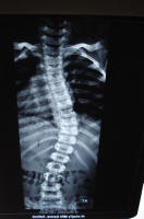 
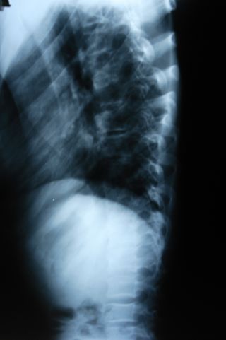

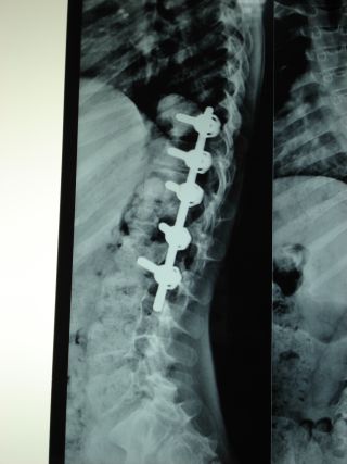
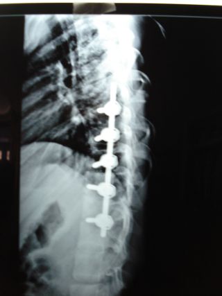
A 12 years old girl with king type II
curve with a progressive deformity.The curve is structural ,with
reasonable amount of flexibility on stress films with a huge rib
hump despite 40 degree curve in stress film. To achieve anterior
fusion and to prevent any possibility of crank shaft phenomenon
occurring and to avoid damaging the lumbar extensor mechanism
selective fusion of the lower thoracic curve was performed. The
upper thoracic curve showed 5 degree decompensation but the
overall sagital and coronal correction was excellent in the post
operative radiogram. The rib hump was reduced drastically. The
decompensation of upper thoracic curve disappeared on follow
up.
Observation
and Results:
20 children with
adolescent idiopathic scoliosis underwent anterior spinal fusion
with single solid rigid rod anterior instrumentation. All were
available for follow up. The average follow up was 2 years,
ranged from half year to 4.5 years. The deformity was
progressive in all patients preoperatively, and documented
radiologically. The average major thoracic curve was 70°, with
range of 55-85°, which was corrected to 12° post operatively,
with the correction rate of 83%. The average secondary
thoracolumbar scoliosis was 40° (20-60°), corrected to 10° post
operatively, with 75% correction. The overall correction was
80%.The average pre operative compensatory curves in the
thoracolumbar region 30° (range 20-40°). The lumbar secondary
curve in thoracic major curve is 37° (range 20-44°). After
correction it was 15° and 17° respectively. In one patient
there was decompensation of secondary thoraco-lumbar curve to
15° compared to the preoperative film. It was treated with
brace and on follow up the curve improved to 8°. The
flexibility of the primary thoracic curve was 68% on stress film
in by Cobbs method. The flexibility of thoracolumbar curve was
80%, in thoracic primary curves. In seven patients distal end
vertebra could be saved by pre operative evaluation using stress
films. The sagital alignment preoperatively in the thoracic
region was thoracic kyphosis of 0-18° preoperatively, which
improved to 12° postoperatively. The lumbar lordosis was 52°
preoperatively (range 24-75°) improved to 43° post operatively.
The thoracic and lumbar sagital alignment improved in all
patients post operatively. The average coronal plane lateral
trunk shift was 15 mm (range 0-30 mm), improved to 7 mm after
surgery. On follow up, the trunk shift was 7.5 mm. One patient
had a coronal plane decompensation in thoracolumbar spine by 5
mm. Sagital plane trunk offset improved from 20mm (range 0-40
mm) before surgery to 8 mm (range 0-16 mm) in follow up. There
was no sagital plane decompensation in any patient.
At 2 years
follow up, there was a loss of correction of 0.5°. End vertebra
and stable vertebra tilt improved by 85% and 88% respectively.
The tangential rib hump angle was corrected by 75% (overall).
Apical vertebra rotation was corrected by 68% by derotation
maneuver after rib hump resection. Only autogenic bone grafts
were used in this study. Average blood loss in this study was
1200ml. The average operation time was 210 Minutes. No
neurological or vascular complications were noted intra
operatively or post operatively. Stagnara wake up test was done
in all patients after instrumentation. Fusion could be achieved
at all levels in the latest follow up. No implant failure was
present till the latest follow up. Minor complications like UTI
in 5 patients, paralytic ileus in 10 patients, excess chest
drainage in 1 patient was noted. Intercoastal chest drainage
was used in all patients for 48 patients. In the patient in
whom, there was thoracolumbar decompensation, the trunk shift
was minimal. This decompensation was due to very high degree of
correction of the primary curve. This was treated by post
operative bracing and on follow up, the curve improved.
Scoliosis
research society instrument (16) questionnaire was done in all
patients. All patients were available for follow up. Average
satisfactory score was 4.3/5. Self image score was 4.4/5.
Functional score was 4.5/5. Mental health score was 4.2/5.
Overall total score was 84%. All patients were satisfied with
the results.
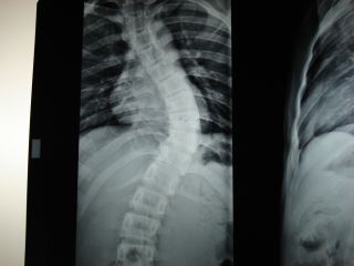
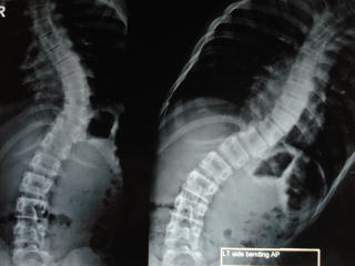
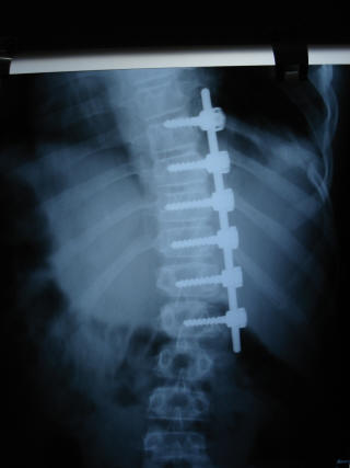
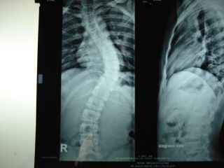
A 13 years old girl with type 2 king
curve showing fairly good flexibility in stress films was
treated with anterior instrumentation and fusion with excellent
post operative post operative correction in both sagital and
coronal plane
Discussion:
The introduction
of anterior scoliosis surgery by Dwyer et all (1) provided a
powerful innovative method of deformity correction in the
spine. Anterior correction of scoliosis in adolescent
idiopathic scoliosis group in the thoraco lumbar and lumbar
spine has already been reported with good results(17,18,19,20)
with less implant problems, more rapid healing and lower
pseudoarthrosis rate than posterior instrumentation and fusion,
even with vertebral screws. Kaneda et all reported Kaneda
Anterior Spinal Stabilization System (KASS), to correct the
deformity with a rod rotation maneuver
(10,11). The fusion segments were in the area of Cobbs
angle. Number of segments fused was less than that of Posterior
fusion. Even though thoracic curves are severe, reports of
thoraco-lumbar and lumbar curves treated by anterior
instrumentation is prevalent in literature.(11) We could find
only scanty reference in English literature regarding correction
by anterior instrumentation in adolescent idiopathic scoliosis
(10).
Thoracic spine
is connected to the ribs by the costo transverse and rib head
articulation of the vertebra. This forms a rigid anatomical
structure compared to thoraco lumbar or lumabar spine. The
thoracic scoliosis is more rigid than thoracolumbar or lumbar
curves for the same reasons (21).
Rib head resection with rod rotation maneouver makes the
thoracic spine less rigid (10).
Oda et all (22) reported
biomechanical role of costo vertebral articulation and the rib
cages in the stability of thoracic spine and concluded that
these were the significant stabilizers in lateral bending and
axial rotation. Anterior release and rib head resection makes
the curve less rigid(10). So a high rate of correction can be
obtained with shorter segment fusion, compared to posterior
procedure, by rib head resection, derotation after anterior
release. Dickson et all (23)
stressed that the axial plane rotational deformity has got its
own importance when attempting sagital and coronal plane
correction. Theoretically 3 D correction should solve all the
problems in scoliosis, but clinically the unsightly rib hump
causes the major cosmetic concern. Anterior thoracoplasty is a
useful adjunct to anterior instrumentation. Kaneda et all
reported good results without any incidence of implant failure,
with no evidence of pseudo arthrosis even with single screw
single rod system (10,11). Twin
et all reported good results in another study with single rod
anterior instrumentation (24).
One of the main points assessed in this manuscript is to address
the sagital plane effect of adolescent idiopathic scoliosis in
thoracic spine. The authors have noted, the maintenance of
normal thoracic kyphosis and lumbar lordosis on follow up.
Adolescent idiopathic scoliosis is mainly a hypo kyphotic
scoliosis and kyphogenic effect of thoracic anterior
instrumentation has resulted in normal kyphosis in the thoracic
spine. Also maintenance of normal thoracic kyphosis has
resulted in maintenance of normal lumbar lordosis. Since only
thoracic major curves were selected for this study the sagital
plane correct ion in span of instrumented curve is to be
stressed.
Discectemy with
end plate removal and bone graft has resulted in 100% fusion.
It has been reported that (11)
dual rod system with two screws are more rigid than single rod
system. However all dual rod systems require placement of 2
screws in each segment. This many be impossible in smaller
thoracic vertebrae in a child. There is increased risk of canal
penetration in 2 screw systems against in 1 screw system. In 2
screw system, staple is usually a must, which needs ligation of
segmental vessel at every level. In single screw system,
selected segmental vessels may be saved from ligation. This
avoids the danger of ischemic cord necrosis, especially when the
vessel to be ligated is the artery of Adam Kewicz, which gives
the major blood supply to the spinal cord. The present study
has shown 100% fusion rate with single solid rigid rod anterior
instrumentation and the correction is good as dual rod system
with disc excision and fusion. The present study also shows
that the technique when compared to standard posterior fusion
techniques has led to selection of less fusion segments (the
distal most vertebra can be saved). This may allow normal
function of the spine with mobility. The options in adolescent
idiopathic scoliosis are stand alone anterior procedure with
fusion, stand alone posterior procedure or combined procedure.
Anterior surgery is a viable option, but the case selection
should be accurate. The compensatory curve should be flexible.
In a study by
Brodner et all (25) the patient
group with thoracolumbar anterior correction for the average 40
° curve, the segments fused were 5.6. This resulted in half the
levels to be fused when compared to posterior fusion. In this
study the average segments fused was 5 for an average curve of
70 degrees. This is probably because of additional derotation
facilitated by rib head resection in this study. The current
study also showed sagital correction of thoracic curve from 9
degrees of kyphosis to 12 degress of kyphosis. The lubmbar
lordosis improved from 52 degrees to 43 degrees. The average
coronal plane curve correction was by 83% in the current study.
Harrington rod with posterior instrumentation needs fusion of
more segments with ensuing back pain due to degeneration
(26). This can be prevented by
anterior surgery. In the reported series of anterior
instrumentation, the correction rate was 47%(11), 58%(18)(20)
and 71%. Another series (27) included
6.8 segments in the thoracic spine. Average 6 ° loss of
correction in the frontal and 8 ° correction in the sagital
plane has been reported in single rod system
(28). Kaneda et all reported 1.2 ° coronal and 1 °
saggital plane loss of correction in the thoracic spine
(10). (But was with dual rod
system in the lower thoracic and lumbar region) The correction
loss in our patient was loss of 1° in the coronal and 0.5° in
the sagital plane(in to hypokyphosis) respectively. This is
probably due to newer segmental single rod systems used in this
study. This may confirm the usefulness of single screw single
rod system in the thoracic spine anteriorly. Suggestion of
athletic activity after scoliosis surgery is a matter of debate
(29). The authors believe that the anterior surgery
offers the best chance of return to athletic activities because
of more biologic nature of the procedure with minimum segments
fused, correction at the site of deformity with preservation of
mobility. Even at this point of writing this paper we are
following up these patients and a longer term follow up may
probably tell us the further functional benefits of anterior
short segment correction in thoracic scoliosis.
In this study
sub periosteal rib head resection and rod rotation has been done
in 20 patients. In these patients the correction rate of apical
vertebral rotation was 62%. Pre operative sagital deformity
ranged from 5° lordosis to 30° kyphosis. Post operatively it is
corrected to 12° kyphosis, which stresses the effectiveness of
anterior procedure in maintaining sagital plane alignment.
External support with Knight Tailors brace was used until
fusion. This is especially useful to control the secondary
curves. This study included 4 patients with Type II curves.
One patient had thoracolumbar trunk shift as a part of
decompensation, which was managed with brace and later
improved. Bridewell et all studied 82 patients with adolescent
idiopathic scoliosis (right thoracic scoliosis) treated by CD
instrumentation using posterior derotation maneuver
(30). They found 8 patients with
gross decompensation on follow up. Majority were type II
curves. They stated that in type II curves, too much correction
of the primary thoracic curve beyond the flexibility of the
lumbar curve may produce decompensation. Schilenzke
(31) et all compared the
radiographic outcome of Harrington rod and CD instrumentation.
They stated that rotation was unchanged in Harrington group.
But rotation was increased in CD group in comparison to pre
operative assessment. This indicates that derotation maneuver
transfers the rotation to the un instrumented lumbar curve. In
anterior correction of scoliosis also the same principle
applies. So it could cause decompensation if the lumbar
compensatory curve if it has rotary elements or if the thoracic
curve is too rigid in stress films. Over correction of thoracic
curve should be prevented so that decompensation will not
occur. In type III and IV curves, thoracic correction is enough
for overall curve correction.
Conclusion:
Anterior
correction of thoracic curve in adolescent idiopathic scoliosis
leads to excellent curve correction in the frontal and saggital
plane, by fusing the thoracic major curve. There is no
incidence of excess kyphosis, implant failure or gross loss of
correction or pseudoarthrosis on follow up. Over correction of
the type II curves may lead to decompensation. Rib head
resection facilitates derotation maneuver. Anterior
thoracoplasty is a usual adjunct in anterior scoliosis surgery.
Spontaneous correction of the compensatory curves can occur.
This procedure is not kyphogenic sagital plane correction was
excellent. Usefulness of this more physiological procedure is
stressed in the current study, even though there are only scanty
reports available in English literature.
Reference :
-
Dwyer AF, Newton NC, Sherwood AA. An anterior approach to
scoliosis. Clin Orthop 1969;62:192.
-
Harrington PR. Treatment of scoliosis: Correction and
internal fixation by spine instrumentation. J Bone Joint Surg
Am 1962;44:591-610
-
Mielke CH, Lonstein JE, Denis F,
Vandenbrink K, Winter RB. Surgical treatment of
adolescent idiopathic scoliosis: A comparative analysis. J Bone
Joint Surg Am 1989;71;1170-7.
-
Fitch RD, Turi M, Bowman BE, et al. Comparison of
Cotrel-Dubousset and Harrington rod instrumentation in
idiopathic scoliosis. J Pediatr Orthop 1990;10:44-7.
-
Schufilebarger HL, Clark CE.
Cotrel-Dubousset instrumentation. Orthopedics
1988;11:1435-40.
-
Horton WC, Holt RT, Johnson JR, Leatherman KD. Zielke
instrumentation in idiopathic scoliosis: Late effects and
minimizing complications. Spine 1988;13:1145-9.
-
Moskowitz A, Trommanhauser S. Surgical and clinical
results of scoliosis surgery using Zielke instrumentation.
Spine 1993;18:2444-51
-
Trammel TR, Benedict F, Reed D. Anterior spine fusion
using Zielke instrumentation for adult thoracolumbar and lumbar
scoliosis. Spine 1991;16:307-16.
-
Wojcik AS, Webb JK, Burwell RG. An analysis of the
effect of the Zielke operation on S-shaped curves in idiopathic
scoliosis: The use of EVAs showing that correction of the
thoracic curve occurs in its lower part: Significance of the
thoracolumbar spinal segment. Spine 1989;14:625-31.
-
Kaneda K, Shono Y, Satoh S, et al.
Anterior correction of thoracic scoliosis with Kaneda anterior
spinal system. A preliminary report. Spine 1997;22:1358-68.
-
Kaneda K, Shono Y, Satoh S, et al. New
anterior instrumentation for the management of thoracolumbar and
lumbar scoliosis: application of the Kaneda two-rod system.
Spine 1996;21:1250-62.
-
Dunn HK. Anterior stabilization of thoracolumbar injuries. Clin
Orthop 1984;189:116-24.
-
Hopf CG, Doz P, Eysel P, et al. Operative
treatment of Scoliosis with Cotrel-Dubousset-Hopf
instrumentation. Spine 1997;22:618-27.
-
Betz RR, Harms J, Clements DH, et al.
Comparison of anterior and posterior instrumentation for
correction of adolescent thoracic scoliosis. Spine
1999;24:225-39.
-
King HA,Moe JH,Bradford DS,WinterRB.Selection of fusion levels
in thoracic idiopathic scoliosis. J Bone and Joint Surg
Am1983;65:1302-13.
-
Asher MA, Lai SM, Burton DC. Further development and validation
of the Scoliosis Research Society (SRS) outcome instrument.
Spine 2000;18:2381-6.
-
Zielke K. Ventral duration spondylodesis. Results of treatment
of cases of idiopathic scoliosis. Z Orthop Ihre Grenzgeb
1982;120:320-9.
-
Bernstein RM, Hall JE. Solid rod short segment anterior fusion
in thoracolumbar scoliosis. J Pediatr Orthop B 1998;7:124-31.
-
Majd ME, Castro FP, Holt RT. Anterior fusion for idiopathic
scoliosis. Spine 2000;25:696-702..
-
Sweet FA, Lenke LG, Bridwell KH, et al.
Prospective radiographic and clinical outcomes and complications
of single solid rod instrumented anterior spinal fusion in
adolescent idiopathic scoliosis. Spine 2001;26:1956-65.
-
Panjabi MM, Hausfeld JN, White AA. Abiomechanical study of the
ligamentous stability of the thoracic spine in man. Acta Orthop
Scand 1981;52:315-26.
-
Oda I, Abumi K, Lu D, Shono Y, Kaneda K. Biomechanical role of
the posterior elements, costovertebral joints, and rib cage in
the stabiloity of the thoracic spine. Spine 1996;21:1423-9
-
Dickson RA, Lawton JO, Archer IA, Butt WP. The pathogenesis of
idiopathic scoliosis: Biplanar spinal asymmetry. J Bone Joint
Surg (Br) 1984;66:8-15.
-
Turi M, JohnsonCE, Richards SB. Anterior correction of
idiopathic scoliosis using ISRH instrumentation. Spine
1993;18:417-22.
-
Brodner, Wolfram, Mun Yue, Wai, Hans B Moller, Kelly Hendricks
et al. Short segment Bone-on-bone instrumentation for single
curve idiopathic scoliosis. 1: 2003 Oct
15;28(20):224-33--------2003;28:224-233.
-
Conolly PJ, Von Schroeder HP, Johnson GE, et al. Adolescent
idiopathic scoliosis. Long term effect of instrumentation
extending to the lumbar spine. J Bone Joint Surg Am
1995;77:1210-16.
-
Rhee JM, Bridwell KH, Won DS, et al.
Sagittal plane analysis of adolescent idiopathic scoliosis. The
effect of anterior versus posterior instrumentation. Spine
2002;27:2350-6.
-
Ouellet JA, johston CE. Effect of grafting technique on the
maintance of coronal and sagittal correction in anterior
treatment of scoliosis. Spine 2002;27:2129-35
-
Rubery PT, Bradford DS. Athletic activity after spine surgery
in children and adolescents. Results of a survey. Spine
2002;27:423-7.
-
Bridwell K H,Mc Allister JW,Betz RR,Hus G,Clancy
M,SchoeneckerPL.Coronal decompensation produced by
Cotrel-Dubousset Derotation maneuver for idiopathic right
scoliosis.Spine 1991:16:769-77.
-
Schlenzka D,Poussa m,Muschik M.Opearative treatment of
adolescent idiopathic scoliosis:harrington DTT versus Cotrel-
doubousset instrumentation.J spinal disord1991;4:188-92.
|
|
This is a peer reviewed paper Please cite as
:
Gopinathan P : Short Segment Anterior Correction Of
Thoracic Scoliosis With Single Solid Rigid Rods.( In Adolescent
Idiopathic Scoliosis)
J.Orthopaedics 2008;5(2)e11
URL:
http://www.jortho.org/2008/5/2/e11 |
|
|

