|
Abstract:
Background: Treatment of the anterior acetabular fractures is
known to have worse results than other fracture types. The
operative results of the anterior acetabular fracture, however,
have not been well documented and the literature concerning them
is sparse. To determine the results of operative treatment for
anterior column acetabular fractures, we did a prospective
cohort study of 17 cases of anterior column fractures. Methods:
A total of 17 patients had anterior column fractures, with an
average follow-up period of 24 months; 1 anterior wall, 3
anterior column, 8 anterior fracture with posterior
hemitransverse and 5 anterior wall with anterior column
fractures. There were 15 men and 2 women, with a mean age of
36.6 years. The surgical approaches used were ilioinguinal
approach used in 14 cases and extended iliofemoral approach used
in 3 patients with delayed presentation. Postoperative
radiographic results were evaluated by Matta’s criteria. Final
clinical results were evaluated by a Harris Hip scoring system
Results: All of the fractures healed. Postoperative radiology
revealed 8 cases of anatomical reduction, 8 cases of imperfect
reduction, and 1 cases of poor reduction. According to the
clinical results, 16 patients had satisfactory results (11
excellent, 5 good), and 1 had poor result. Regarding
complications, there were 1 case of traumatic osteoarthrosis and
1 of heterotopic ossification. The patients with an anatomical
reduction had a higher satisfactory result rate. Poor reduction
intra operatively seemed to have an adverse influence on the
postoperative radiologic result as well as a correlation with
the development of traumatic arthritis.
Conclusion: Anterior acetabular fractures have comparatively
higher rates of imperfect reduction and may have a tendency
toward traumatic osteoarthritis; however a longer follow up
study is essential.
J.Orthopaedics 2009;6(4)e7
Keywords:
anterior acetabulum fractures; surgical management
Introduction:
Acetabular fractures are still and enigma and pose a major
challenge to treating orthopaedic surgeon1. There is
increase in the incidence of high velocity trauma resulting in
complicated acetabulum fractures due to modernization2.
The surgical treatment of acetabular fractures is a complex area
that is being continually refined. Presently open anatomical
reduction of the articular surface combined with rigid internal
fixation and early mobilization is the standard treatment for
these injuries3,4,5,6,7,8. The surgical management of
these fractures involves a definite learning curve, probably
best documented in a report by Matta and Merritt3 of
the first 100 acetabular fractures treated operatively by Matta.
Grouping the surgical reductions chronologically in groups of 20
clearly demonstrated that experience improved the ability to
avoid unsatisfactory reductions and to perform anatomical
reductions.
Problems like complex anatomy, difficult surgical approaches,
perfect anatomic reduction necessary as major weight bearing
joint, less space for operative maneuverability, comminution,
and delayed presentation pose a challenge for the operating
surgeon. Long term results, no matter which operative approach
is used or fracture type is involved, are directly related to
the quality of fracture reduction achieved6,9 . This
point was first underlined by the long term follow up studies of
Letournel and Matta in which they demonstrated that the
fractures reduced to within 1mm of residual articular
displacement have less incidence of posttraumatic arthritis and
have a more durable and long-lasting functional hip joint than
those fractures with 1 to 3 mm of residual displacement5,10.
These series of operative management are considered as the gold
standard in operative management of acetabulum fractures.
Pioneering work was done by Letournal and Judet in 1964 to
systematically classify the acetabular fractures and to develop
a logical line of thinking regarding management of these
fractures11. Letournal and Judet conceived acetabulum
to be made of two columns. Anterior column from below the
sacroiliac joint to the ischial tuberosity and posterior column
from superior iliac crest to pubic symphysis with both columns
attached to the sacrum by thick strut of bone lying above
greater sciatic notch and called sciatic buttress. Patients with
associated fracture types according to the Letournel
classification and those with injuries to the anterior wall and
posterior column are most likely to have a poor functional
outcome12,5. The anterior fractures are rare and
literature about
the operative results of anterior acetabular fracture is sparse.
Here, we analysed our results regarding the surgical treatment
of displaced anterior acetabular fractures with or without a
minimal posterior involvement.
Materials
and Methods:
This is a prospective cohort study of 17
cases of anterior acetabular fractures treated operatively
between 2004 and 2006. The patients with anterior wall, anterior
column, anterior fracture with posterior hemitransverse and
anterior wall with anterior column fractures were included in
the study taking into account Letournel’s classification5,13.
The follow-up was for a minimum of 2 years. 6 patients had
isolated acetabular fracture, and 11 had other associated
fractures. All patients gave an informed consent to participate
in the study and were prospectively followed up.
The patients with anterior wall, anterior column, anterior
fracture with posterior hemitransverse and anterior wall with
anterior column fractures were included in the study taking into
account Letournel’s classification13. The indications
for surgery were fractures of the anterior wall and/or column
that are characterized by intraarticular gaps or steps of > 3 mm
in the area of the main weight-bearing dome of the acetabulum,
fractures complicated by subluxation or dislocation of the
femoral head, an intraarticular fragment making the joint
incongruous and interfering with joint movement and roof arc
angle < 45deg. The patients with revision surgeries were
excluded. All patients were initially stabilized
hemodynamically and then anteroposterior (AP) and Judet views
were taken. A CT scan with a 3D reconstruction was obtained
preoperatively in all patients. Patients were immobilized in a
skeletal traction after the radiographs were taken. The
fractures were classified according to the Letournel and Judet
classification.
All the patients were operated within 4 weeks of trauma. The
ilioinguinal approach was used in most patients while extended
iliofemoral approach was used in cases with delayed presentation
> 3 weeks. The ilioinguinal approach was performed with the
patient in a supine position with a slightly elevated
ipsilateral half of pelvis and the hip and knee flexed to 30 to
40 degrees to relax the neurovascular structures under the
inguinal ligament. Intraoperative fluoroscopy was used to assess
reductions.
Immediate postoperatively AP views and Judet views were taken.
Post operatively all patients were immediately mobilized non
weight bearing with the help of crutches or walker except who
had polytrauma. All patients were reviewed clinically and
radiologically at 3, 6, 12 and 18 months. After that they were
reviewed every 12 months.
All operations were
performed jointly by the 2 senior authors. Cefuroxime was used
as prophylactic antibiotic. Postoperatively, 75mg of
Indomethacin in 3 divided doses daily was given for 6 weeks
for
prophylaxis against heterotopic
ossification14.
Follow-up included ongoing evaluation with radiographic films
and assessment of range of motion of hip joints, degree of pain
using the Visual Analogue Scale (VAS), and the degree of
ambulation. The outcome of patients was evaluated by the Harris
Hip Scores (HHS) and Visual Analogue pain Scale. Postoperative
radiographic results were evaluated by Matta’s10
criteria (anatomic reduction <1mm; imperfect 1–3mm; poor >3mm).
The patients were at the latest follow-up graded as per
ambulation status and shortening.
The presence of ectopic bone, sclerosis, spur formation of
femoral head, congruence of the femoral head with acetabulum,
signs of degeneration of the femoral head and acetabulum were
assessed from the radiographs. Heterotopic ossification was
evaluated and graded according to the classification of Brooker
et al 15
Results :
There were 15 males and 2 female patients with average age of
36.6 years (range, 17-66 years). The mechanisms of injury were
fall from a two-wheeler in 8 patients, road accidents in 6, and
3 patients had a fall from height. 6 were left sided fractures
while 11 were right sided. We did not have any patients with
bilateral acetabular fractures.
The fractures were classified according to Judet and Letournel
with 1 anterior wall, 3 anterior column, 8 anterior fracture
with posterior hemitransverse and 5 anterior wall with anterior
column fractures. None of the cases had any compound fractures.
One patient had sciatic nerve palsy preoperatively. All patients
had a displaced fracture of acetabulum with intraarticular step
more than 2 mm in all. The average roof arc angle as measured on
AP view, obturator oblique view and iliac oblique view are 17.29
degrees, 20.24 degrees and 19.77 degrees respectively.
Out of the 17, six were polytrauma cases with 1 patient with a
head injury and 1 with chest injury. There were no associated
spinal injuries. 6 cases had associated ipsilateral lower
extremity fractures and 4 had associated upper extremity
fracture. 3 patients also had concomitant pelvic ring injuries
and 1 had a femoral neck fracture. 2 patients had Moralle-
Lavalle lesions, both of which resolved spontaneously. None of
the patient had any associated bladder or urethral injuries. 4
cases presented with central hip dislocations along with
anterior columnar fractures which were reduced with lateral
skeletal traction. We had no incidence of anterior or posterior
dislocations.
The average injury- operative interval was 8 days (range 1-28
days). The ilioinguinal approach was used in 14 cases and
extended iliofemoral approach was used in 3 patients with
delayed presentation. 5 patients had comminution of which 3 were
anterior fracture with posterior hemitransverse and 2 were
anterior column with a quadrilateral plate fracture.
The average operating time including the positioning of patient
was 158 min (range 90- 320). The average blood loss was 515 ml
(range 350-850 ml) and blood transfusions were required in 8
patients.
The quality of reduction was measured postoperatively on
radiographs and was anatomic in 8, imperfect in 8 and poor in 1
case as per grading by Matta et al.
The average Visual Analogue Pain Score at the end of 3 months
was 6.13 (range 5-8), at the end of 6 months was 3.94 (range
3-5), at the end of 1 year was 3.19(range 2-5) and at the end of
2 years was 2(range 1-5).The average HHS at 3 months was 69, at
6 months was 77, 1 yrs was 83.7, and at 2 yrs was 90.05.
According to the clinical results, 16 patients had satisfactory
results (11 excellent, 5 good), and 1 had poor result.
Heterotopic ossification developed in 1 patient who had Grade II
according to Booker’s classification. He had no functional
impairment due to heterotopic ossification.
Osteonecrosis was seen in none of the cases. 1 patient had
post-traumatic osteoarthrosis of the hip. As expected, he had
non anatomic reduction of intraarticular fragments with a poor
reduction as per Matta’s grading . He developed a shortening of
1.5 cms. Another patient of associated polytrauma had 1 cm of
post-operative shortening.
Additional complications included a superficial wound infection
and a hematoma which healed with oral antibiotics without
further problems. None of the cases had any intraoperative or
postoperative neurovascular complication although one case had
preoperative partial sciatic nerve palsy which did not recover
fully at the final follow-up.
At the final follow-up 14 patients were walking independently of
any walking aid, 2 were walking full weight bearing with help of
a walking stick and 1 patient who had had an above knee
amputation walking with an artificial prosthesis. Figure 1 to 4
shows two cases of our series with pre operative and final
radiographs.
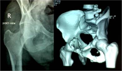
Figure 1: Twenty three year old male having right
anterior column fracture as seen on Judet view and 3-D CT scan
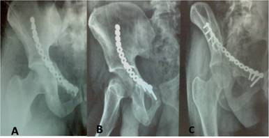
Figure 2: same patient as in fig 1 treated by
ilioinguinal approach using reconstruction plate[A]. B and C
show 30 months follow up with good union
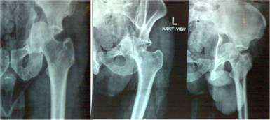
Figure 3: thirty four year male with anterior column and
posterior hemitransverse fracture as seen on antero-posterior
and Judet view radiographs.
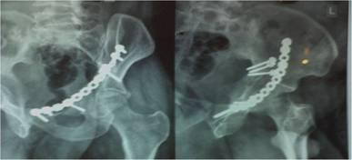
Figure 4: Same patient as shown in Figure 3 treated with
anterior plating with good union at 36 months follow up.
Discussion :
To our knowledge this is the first prospective series studying
the anterior acetabular fractures in English literature. 16 of
17 patients operated upon in our series had excellent or good
result at a early follow up of 2 years and these results were
comparable to other authors.
According to a recent metaanalysis13 the anterior
fractures constitute about 10.2% of the total acetabular
fractures (this included the anterior wall, anterior column and
anterior with posterior hemi-transverse). A road traffic
accident was the causative mechanism in 80.5% of patients, 10.7%
had falls and in 8.8% other causes were stated. In our series
there were 14 cases of road traffic accident (82.3%) and three
cases of fall from height which is almost similar to the
meta-analysis data. Pre operative sciatic nerve injury was noted
in 16.4% in the above article while we had only one case (5.8%)
of sciatic nerve injury. This may be because most of the sciatic
nerve injuries were noted in posterior fractures especially
those with posterior dislocation and our series had no cases of
posterior dislocation.
The goal of acetabular fracture treatment is to have a hip with
good long-term function and the avoidance of posttraumatic
osteoarthritis7,10,16. Letournel and Judet17
introduced the operative concepts of open reduction and internal
fixation for acetabular fractures. The extent of influence the
initial fracture pattern has on clinical outcome of acetabular
fractures has been studied18,19,20. The radiographic
roof arc angle is originally used to determine whether a
fracture line crosses the weight-bearing dome21. It
is also important to know the extent of the fracture when trying
to predict clinical outcome. In our series all cases were
selected according to the indications for operative treatment as
stated by Matta et al thus this bias was eliminated. There were
however 3 cases with comminution but there effect on final
outcome cannot be pointed out as none of the cases with fair or
poor results were having pre operative comminution.
Over the years the use of ilioinguinal approach was emphasized
because of the good results it usually provided including non
development of heterotopic ossification and quick
rehabilitation. Giannoudis et al13 stated that
incidence of HO is overall 25.6% and differs with surgical
approach with highest incidence of 23.6% for iliofemoral
approach and incidence of 1.6% for ilioinguinal approach. Chiu
et al22 reported incidence of 5.6% with ilioinguinal
approach and 66.7% in cases operated by iliofemoral approach. In
our series we had only one case of booker grade II HO in a case
operated by the iliofemoral approach. However since we used this
approach in cases with delayed presentation only, this may also
be a confounding factor in development of HO. This case in our
series had good result and no functional limitation that can be
attributed to the HO. We had no case of HO in cases operated
with ilioinguinal approach thus agreeing with the literature
about the low incidence of Ho, however, reduction of the
posterior column can be a problem with this approach, especially
of there is also a rotation of the posterior column.
A minimum follow up of 2 years was chosen as an acetabular
fracture is intraarticular and osteoarthrosis will usually
develop within the first 2 years. No major changes were found in
clinical, functional and radiographic results between 1 to 2
years postoperatively. It appears that if the result is
clinically and radiologically good or excellent and stable from
the time of operation, without any signs of degenerative disease
after 1 year, the long term outcome will not usually change
however a long term study will be essential to study the
validity of this statement. Traumatic osteoarthrosis is one of
the worst complications of acetabular fractures, and many
clinical studies have found that incongruent reduction of an
acetabular fracture can lead to poor functional outcome because
of posttraumatic arthrosis5,16. Analysis of
literature states that the postoperative reduction was recorded
as being satisfactory, with less than 2 mm of displacement, in
85.6% of fractures13. In our study however there were
8 cases with imperfect reduction and one case with poor
reduction i.e. a total of 9 out of 17 (53%) with non anatomical
reduction. This percentage for anterior acetabular fractures is
significantly more than what is stated in literature for all
acetabular fractures. Giannoudis et al13 also noted
that if the reduction was satisfactory (≤ 2 mm), the incidence
of osteoarthrosis was 13.2% and if the reduction was not
satisfactory (> 2 mm), it increased to 43.5%. In this study with
a relatively short follow-up, 1 of 17 patients (5.8%) developed
osteoarthritis. As anticipated, this patient had not achieved
anatomical reduction. However a longer follow-up will be
required to study the correlation between imperfect reduction
and development of osteoarthrosis in our series although we
believe that anatomical reduction of the weight-bearing dome of
the acetabulum should be achieved to minimize the incidence of
posttraumatic arthritis.
There are several limitations to the present study. The
incidence of anterior acetabular fractures is relatively lower
than that of posterior fractures, so the study is underpowered
and the number of patients may be insufficient to draw concrete
conclusions. However since the study is still ongoing we will be
enrolling more patients and follow-up time targeted as minimal 5
years so as to present a comprehensive study of these anterior
acetabular fractures.
Conclusion:
The surgical outcome depends on many factors; the ability of the
surgeon to classify the fracture; choose the appropriate
approach; to have adequate and proper instruments, theatre
facilities and to employ a proper surgical technique so as to
get a near anatomic reduction. Inspite of clearing these hurdles
there are other factors that are not in surgeons control and can
give a poor outcome like late presentation, gross comminution,
and osteoporosis. Present study indicates that anterior
acetabular fractures have comparatively higher rates of
imperfect reduction and may have a tendency toward traumatic
osteoarthritis, however a longer follow up and an appropriately
powered study is essential to make other conclusions regarding
these fractures.
Reference :
-
Tile M, Helfet D,
Kellam J. Fractures
of the Pelvis and Acetabulum. Baltimore. Lippincott Williams
& Wilkins; 3rd edition, 2003.
-
Gupta RK, Singh H, Dev B, Kansay R, Gupta P, Garg S. Results
of operative treatment of acetabular fractures from the Third
World--how local factors affect the outcome. Int Orthop. 2009
Apr;33(2):347-52.
-
Matta JM, Merritt PO.: Displaced acetabular fractures. Clin
Orthop Relat Res. 1988 May;(230):83-97.
-
Letournel E.: Acetabulum fractures: classification and
management. Clin Orthop 1980; 151:81–106.
-
Letournel E, Judet R. Fractures of the acetabulum. 2nd ed. New
York: Springer; 1993. p. 63–6, 565–8.
-
Matta JM, Mehne DK, Roffi R (1986) Fractures of the acetabulum:
early results of a prospective study. Clin Orthop Relat Res
205:241–250.
-
Matta JM, Anderson LM, Epstein HC, Hendricks P. Fractures of
the acetabulum. A retrospective analysis. Clin Orthop
1986;205: 230–40.
-
Matta JM, Merritt PO. Displaced acetabular fractures. Clin
Orthop 1988;230:83-97.
-
Brueton RN (1993) A review of 40 acetabular fractures. The
importance of early surgery. Injury 24(3):171–174.
-
Matta JM. Fractures of the acetabulum: accuracy of reduction
and clinical results in patients managed operatively within
three weeks after the injury. J Bone Joint Surg Am
1996;78:1632–45.
-
Judet R, Judet J, Letournel E. Fractures of the acetabulum:
Classification and surgical approaches for open reduction. J
Bone Joint Surg Am. 1964;46A:1615-38.
-
Giannoudis PV, Grotz MR, Papakostidis C, Dinopoulos H.
Operative treatment of displaced fractures of the acetabulum.
A meta-analysis. J Bone Joint Surg Br. 2005 Jan;87(1):2-9.
-
E Letournel, Acetabular fractures, Current Orthopaedics
Volume 3, Issue 4, October 1989, Pages 233-243.
-
Burd TA, Lowry KJ, Anglen JO. Indomethacin compared with
localized irradiation for the prevention of heterotopic
ossification following surgical treatment of acetabular
fractures. J Bone Joint Surg [Am] 2001;83-A:1783-8.
-
Brooker AF, Bowerman JW, Robinson RA, Riley LH Jr. Ectopic
ossification following total hip replacement: incidence and a
method of classification. J Bone Joint Surg Am
1973;55:1629–32.
-
Mears DC, Velyvis JH, Chang CP. Displaced acetabular fractures
managed operatively: indicators of outcome. Clin Orthop 2003;
407:173–86.
-
Saterbak AM, Marsh JL, Nepola JV, Brandser EA, Turbett T.
Clinical failure after posterior wall acetabular fractures:
the influence of initial fracture patterns. J Orthop Trauma
2000; 14: 230–7.
-
Moed BR, Carr SE, Gruson KI, Watson JT, Craig JG. Computed
tomography assessment of fractures of the posterior wall of
the acetabulum. J Bone Joint Surg Am 2003;85:512–22.
-
Vrahas MS, Widding KK, Thomas KA. The effects of simulated
transverse, anterior column and posterior column fractures of
the acetabulum on the stability of the hip joint. J Bone Joint
Surg Am 1999; 81:966–74.
-
Mears DC, Velyvis JH, Chang CP. Displaced acetabular fractures
managed operatively: indicators of outcome. Clin Orthop
2003;407:173-86.
-
Heeg M, Oostvogel HJ, Klasen HJ.
Conservative treatment of acetabulum fractures: the role of
the weight- bearing dome and anatomic reduction in the
ultimate results. J Trauma 1987; 27:555– 9.
-
Chiu FY, Chen CM, Lo WH. Surgical treatment of displaced
acetabular fractures: 72 cases followed for 10 (6-14) years.
Injury 2000;31:181-5.
|

