|
ABSTRACT
Background
A short term follow up study of pilon fractures of the ankle treated over a period of 5 years in a teaching institution. Pilon fractures are becoming more and more common because of increased incidence of high velocity injuries. Many are compound fractures, since Tibia is a subcutaneous bone. Outcome of such fractures are generally not satisfactory because of secondary
osteoarthritis, which occur as a sequele of inadequate reconstruction of the joint. 1 mm tilt of Talus changes the weight transmission by 25%. Primary reconstruction with achievement of joint congruence is the only method by which secondary osteoarthritis can be prevented.
Methods
21 patients during the period of 1997-2002 were assessed. Majority were males. 8 of these fractures were compound. Initial management consisted of heamodynamic stabilization and wound debridement in compound fractures. Primary stabilization is done whenever the general conditions were satisfactory. Mode of treatment consisted of biaxial distraction and stabilization by external fixation and limited internal fixation. Primary internal fixation without external fixation was also done in some cases, where internal fixation alone has given good stability to the fracture and joint congruence.
Results
Of the 21 patients studied during the period with an average follow up of 2 ½ years. 13 fractures united with congruent ankle joint, 3 developed
Osteoarthritis, 3 patients had 1-3ºof tilt of the talus, which have painful ankle joint and needs a second surgery. Other patients are still on follow up. 61% of the patients in this series had best outcome.
Discussion
Pilon fractures always cause difficulty in management because of its inherent instability and intraarticular extension. To attain the best outcome needs a perfect reconstruction of articular congruence with best possible alignment. Open reduction is not usually advisable because of severity of communition and loose fragment. Biaxial distraction with limited internal fixation helps a better alignment and articular reconstruction without much trauma to the patient. But in cases where a primary fixation is possible, external fixation can be avoided. Biaxial distraction with limited internal fixation is a good feasible technique in pilon fracture to attain fracture alignment
J.Orthopaedics 2004;1(1)e4
Introduction
Pilon fractures have become a common problem in trauma care due to high energy road traffic accidents and industrial accidents. Pilon fractures due to high velocity trauma results in multiple metaphyseal fragments, displaced intra articular communition and soft tissue injuries; where as Pilon fractures due to low energy injuries, as seen in sporting activities produce spiral fractures of metaphysis with minimal or no displacement of intra-articular fragments(1).
In the past conservative treatment for Pilon fractures of Tibia was preferred due to limited availability of implants and poor outcome of surgical
intervention(1,2). Classic open reduction and fixation with long plates and screws was shown to be associated with a high rate of early soft tissue complications, including skin necrosis, superficial infection, osteomyelitis and even
amputation(2-5).
In high velocity injuries many are compound fractures, since tibia is a subcutaneous bone. Inadequate reconstructions of the joint will result in changes in the weight transmission and early osteoarthritis of the
ankle(4,5,6,7). Primary reconstruction of the joint congruence with minimal disturbance of the soft tissue is the only method by which good function can be
achieved(8,11).
Materials and Methods
Here we present a short term follow up study of pilon fractures of ankle treated over a period of 5 years. 21 patients were treated during the period by 1997-2002 were assessed.
Patients attending to Accident and Emergency department of Calicut medical college, having pilon fractures with intra articular communition and displacement were selected. 21 patients were included in the study. 8 of these fractures were compound. Initial assessment in the A& E department is followed by heamodynamic stabilization and management of life threatening injuries. Other skeletal injuries were treated accordingly. Ankle fracture is assessed with AP and Lateral view X rays. Contra lateral ankles were also X-rayed for comparison. Fractures were classified according to Ruedi and
Allgower.
Wound debridement of the compound fracture is done initially and primary stabilization is done whenever the general condition of the patient were satisfactory.
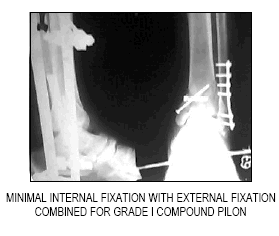
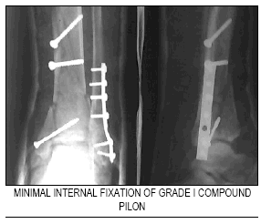
Mode of definite treatment consisted of Bi-axial external fixation with Schanz screws in the upper Tibia and a transverse Steinman pin in the Calcaneum. Manual axial distraction is done using AO Universal Joints and Tubular connecting rods. Indirect reduction of the fracture site was achieved through the soft tissues attachment to the fracture fragments with ligamentotaxis. Tibial plafond fracture site is approached through an anteromedial incision of the ankle. Fracture fragments in the articular surface alone are reduced and internal fixation is achieved with K wires, screws or with plates, depending upon the fracture configuration(9,10). Lateral malleoulus, whenever is fractured is fixed with 1/3 tubular plate or tension band wiring. Limited internal fixation achieved after indirect fracture reduction through ligamentotaxis, usually needs a small incision and wound closure does not pose a problem, unlike in classic internal fixation surgeries.
Primary internal fixation without external fixation was also done in some cases (less severe injuries), where, internal fixation alone has given good stability to the fracture site and congruent joint. We prefer external fixation with bi axial distraction with minimal internal fixation. Bone grafting was done in some cases as a secondary procedure to fill the gap in the metaphysis. Post operative antibiotics are continued for 48 hours parenterally, followed orally till sutures are removed. Analgesics are given post operatively, till suture removal and continued SOS.
External fixator was removed at the end of 6 weeks and gentle ankle motion exercises were begun. Weight bearing was allowed only after the fracture was healed.
Whenever surgery was delayed due to poor general condition of the patients, initial treatment consisted of skeletal traction through a transverse Calcaneal pin, which can be later used for the construction of external fixator frame for Bi-axial
ligamentotaxis.
Evaluation
Patients were assessed for functional, cosmetic and radiological results. Gait was assessed and recorded as either antalgic or non-antalgic. They were assessed for ascend and descend on stairs. Limb length was assessed with the block method. Combined maximum ankle and foot dorsiflexion and plantar flexion were measured and compared with the non injured side. Swelling of the ankle is measured at the level of the medial malleoulus and compared with the opposite side. Wasting of the calf muscles was measured. Neurological examination was performed to assess the sensory abnormalities to light touch and was described according to their distribution of cutaneous nerves.
Results
Of the 21 patients studied during the period with an average follow up of 2 ½ years. 8 of these fractures were compound. Male were involved in 15 cases and 6 were females. In 18 patients there was associated lateral malleoular fracture which needed fixation. Of these 15 patients were treated with 1/3 tubular plate and rest with tension band wiring. In 4 patients, primary internal fixation without external fixation has given good alignment and congruous joint. Post operative infection of the wound was noted in 6 patients. Pin tract of the Calcaneal pin was infected in 8 patients and Tibial pin was infected in 2 patients. All were superficial infection, and subsided with antibiotics. 3 patients needed bone grafting for delayed union. In situ bone grafting at the metaphyseal region is done from the Iliac crest, and the fractures eventually united. 13 fractures united with congruent ankle joint, 3 developed Osteoarthritis, 3 patients had 1-3º tilt of the talus which have painful ankle joint and needed a second surgery. Other patients are still on follow up. 61% of the patients in this series had good outcome.
 |
|
 |
|
Discussion
Pilon fractures always cause difficulty in management because of its inherent instability and intra articular extension. Internal fixation with extension soft tissue dissection often results in unacceptable outcomes. To attain the best results, needs a perfect reconstruction of articular congruence with best possible alignment at the minimum of soft tissue dissection. 38% fractures were compound, as the Tibia is a subcutaneous bone. In 85% of cases, there was an associated Lateral malleoular fracture which needed fixation. In our series, infection, which was superficial, was noted in only 28% cases, which subsided with antibiotics. There was no incidence of deep infection or Osteomyelitis. Good tissue healing was due to minimal soft tissue dissection, as the fracture was reduced indirectly using ligamentotaxis. Good reduction was obtained by bi-axial ligamentotaxis, and 63% patients had congruous articular surface. 15% of the patients developed Osteoarthritis of the ankle due to incongruous joint and Talar tilt. The painful ankle in these patients needs some surgical procedures.
In high velocity injuries producing Pilon fractures, which are associated with sever soft tissue trauma, and communited intra articular displaced fragments, bi-axial external fixation with limited internal fixation, is definitely a good treatment option. Further improvement in fixation devices and more understanding of the biomechanics of the ankle and fracture pattern will contribute to evolve a better treatment plan for this difficult fracture pattern.
Reference
1. Babis GC, Vayanos FD, Papaioannou N, Pentazo Poulose T:- Results of surgical treatment of Tibial Plafond fractures, Clin Orth 341: 99-105, 1997.
2. Bonar SK, March JL: Unilateral external fixation for severe pilon fractures. Foot & Ankle 14: 57-64, 1993.
3. Brumback RJ, Mc Gravey WC: Fractures of tibial plafond; evolving treatment concepts for pilon fractures OCNA Am 26: 273-285, 1992.
4. Burwell HN, Charnley AD: The treatment of displaced fractures of the ankle by rigid internal fixation and early joint movemnts JBJS 47: 634-659, 1965.
5. Helfet DL, Koval K, Pappus J et al: Intra articular pilon fractures of the Tibia, Clin Orth 298: 221-228, 1994.
6. Marsh JL, Bonar S, Nepola VJ et al: Use of an articulated external fixator for fractures of the Tibial plafond. JBJS 77:1498-1509, 1992
7. Tornetta P: Axial compensated tomography of pilon fractures. Clin Orthop 323: 273-276, 1993.
8. Ovadia DN, Beah RK: Fractures of the Tibial plafond JBJS 68: 543-551, 1986.
9. Ruedi & Allgower M: The operative treatment of intra articular fractures of lower end of Tibia. Clin Orth 138: 105-110, 1979.
10. Sanch A, Grujic L, BNyck BC et al: Clinical and funcational outcome of internal fixation of displaced pilon fractures. Clin Orthop 347: 131-137, 1998.
11. Joseph Borvelli, Eric Ellis: Pilon fractures, assessment and treatment OCNA vol 33: 231-248, Jan 2002.
12. Wyrsch B, Mc Ferran MA, Mc Andrew et al: Operative treatment of fractures of Tibial plafond; A randomized prospective study JBJS 78: 1646-1657, 1996.
|

